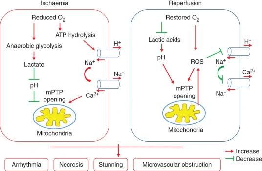
Ischemia/reperfusion (I/R) injury is a complex pathophysiological process that occurs when blood supply is restored to tissues or organs that have been deprived of oxygen and nutrients (ischemia). While the restoration of blood flow (reperfusion) is essential for tissue survival, it paradoxically initiates a cascade of events that can significantly worsen cellular damage and organ dysfunction. This phenomenon is particularly relevant in clinical scenarios such as myocardial infarction, stroke, acute kidney injury, and especially in organ transplantation, where organs undergo mandated periods of ischemia during procurement, preservation, and implantation. Understanding the mechanisms underlying I/R injury is crucial for developing effective strategies to prevent or minimize its devastating consequences.
Explanation of Ischemia/Reperfusion Injury
Ischemia is defined as the insufficient supply of blood and oxygen to an organ or tissue, typically caused by a blockage or constriction of blood vessels. This deprivation leads to cellular hypoxia and an inability to maintain aerobic respiration, forcing cells to switch to less efficient anaerobic glycolysis. This results in reduced ATP production, accumulation of metabolic byproducts (such as lactic acid, causing intracellular acidosis), failure of ion pumps (leading to cellular swelling and electrolyte imbalance, particularly influx of sodium and calcium), and disruption of cellular homeostasis. If prolonged, ischemia leads to irreversible cell damage and necrosis.
Reperfusion is the process of restoring blood flow to the ischemic tissue. While vital for delivering oxygen and nutrients necessary for recovery, the sudden reintroduction of oxygen and circulating immune cells into the compromised tissue environment triggers a complex inflammatory cascade and oxidative burst that can paradoxically exacerbate the injury sustained during the ischemic phase. This ‘reperfusion injury’ is often responsible for a significant portion of the total damage observed. The key events during reperfusion include:
- Rapid generation of reactive oxygen species (ROS).
- Inflammatory cell infiltration and activation (neutrophils, macrophages).
- Activation of the complement system.
- Endothelial dysfunction, leading to increased vascular permeability, capillary leak, and impaired microcirculation (‘no-reflow’ phenomenon).
- Activation of pro-inflammatory signaling pathways (e.g., NF-kB).
- Induction of various cell death pathways, including apoptosis and secondary necrosis.
The interplay of ischemic and reperfusion-related events results in extensive tissue damage, inflammation, and potentially organ failure. The severity of I/R injury is influenced by the duration and severity of ischemia, the type of tissue involved, the temperature during ischemia (cold vs. warm ischemia), and the conditions under which reperfusion occurs.
The Role of Reactive Oxygen Species (ROS) in Ischemia/Reperfusion Injury
Reactive Oxygen Species (ROS) are highly reactive molecules containing oxygen, such as superoxide radical (O₂⁻•), hydrogen peroxide (H₂O₂), and hydroxyl radical (•OH). Under normal physiological conditions, ROS are produced at low levels as a byproduct of metabolic processes (primarily mitochondrial respiration) and play roles in cellular signaling. Cells possess robust antioxidant defense systems (enzymatic, e.g., Superoxide Dismutase – SOD, Catalase, Glutathione Peroxidase; and non-enzymatic, e.g., Glutathione, Vitamins C and E) to neutralize these molecules.
During ischemia/reperfusion, there is a dramatic imbalance between ROS production and antioxidant defense, leading to a state of oxidative stress. ROS generation is significantly amplified during both phases:
- During Ischemia: While oxygen levels are low, some ROS production still occurs due to mitochondrial dysfunction and the activation of enzymes like xanthine oxidase. Ischemia leads to ATP depletion, which causes the accumulation of hypoxanthine (from ATP breakdown). Also, the rise in intracellular calcium activates proteases that convert xanthine dehydrogenase (normally present) into xanthine oxidase.
- During Reperfusion: The sudden reintroduction of oxygen fuels a massive surge in ROS production. This is the major phase of ROS-mediated damage. Key sources include:
- Mitochondria: Reoxygenation leads to rapid electron transfer in the impaired electron transport chain, particularly at Complexes I and III, generating large amounts of O₂⁻•.
- Xanthine Oxidase: With the return of oxygen, the accumulated hypoxanthine is rapidly metabolized by the xanthine oxidase generated during ischemia, producing uric acid and large amounts of O₂⁻• and H₂O₂.
- NADPH Oxidases (NOX): Activated in endothelial cells, phagocytes, and other cell types, NOX enzymes produce O₂⁻⁻•, contributing to inflammation and damage.
- Inflammatory Cells: Infiltrating neutrophils and macrophages generate a “respiratory burst” upon activation, releasing large quantities of ROS and RNS (Reactive Nitrogen Species) via NOX and inducible Nitric Oxide Synthase (iNOS).
The excessive ROS overwhelm the cellular antioxidant defenses. The consequences of this oxidative stress are severe and widespread:
- Lipid Peroxidation: ROS attack polyunsaturated fatty acids in cell membranes, generating lipid peroxides that disrupt membrane structure and function, increasing permeability and leading to cellular and organelle damage.
- Protein Oxidation: ROS can modify amino acids, leading to protein misfolding, aggregation, and loss of enzymatic activity or structural integrity.
- DNA Damage: ROS can cause DNA strand breaks and base modifications, potentially leading to mutations or triggering cell death pathways.
- Activation of Signaling Pathways: ROS act as signaling molecules that activate pro-inflammatory pathways like NF-kB and MAPK pathways, promoting the expression of cytokines, chemokines, and adhesion molecules, thus amplifying the inflammatory response.
Targeting ROS production and enhancing antioxidant defenses is a major strategy in mitigating I/R injury.
The Role of Apoptosis in Ischemia/Reperfusion Injury
Apoptosis, or programmed cell death, is a genetically regulated process essential for development, tissue homeostasis, and removing damaged or infected cells. Unlike necrosis, which is typically a chaotic and inflammatory process resulting from severe, acute injury, apoptosis is characterized by distinct morphological changes (cell shrinkage, chromatin condensation, formation of apoptotic bodies) and biochemical events (activation of caspases). While necrosis is often considered the primary mode of cell death during severe ischemia, apoptosis plays a significant role in I/R injury, particularly during the reperfusion phase and in cells that have sustained sub-lethal damage.
Apoptosis in the context of I/R injury is triggered by multiple converging pathways:
- Mitochondrial Pathway (Intrinsic Pathway): Ischemia and reperfusion cause mitochondrial dysfunction, including loss of membrane potential and increased permeability. This leads to the release of pro-apoptotic factors from the mitochondrial intermembrane space into the cytoplasm, such as cytochrome c. Cytochrome c binds with Apaf-1 and pro-caspase-9 to form the apoptosome, which activates caspase-9 (an initiator caspase). Activated caspase-9 then cleaves and activates executioner caspases (e.g., caspase-3, -6, -7), which dismantle the cell. The balance between pro-apoptotic (e.g., Bax, Bak, Bid) and anti-apoptotic (e.g., Bcl-2, Bcl-XL, Mcl-1) proteins of the Bcl-2 family, often localized to the mitochondria, dictates the sensitivity to this pathway; I/R typically shifts this balance towards apoptosis. Calcium overload during I/R is a potent trigger for mitochondrial dysfunction and the intrinsic pathway.
- Death Receptor Pathway (Extrinsic Pathway): Inflammatory cytokines (e.g., TNF-α) and activation of surface death receptors (e.g., Fas, TNFR1) during reperfusion can activate the extrinsic apoptotic pathway. Ligand binding to these receptors leads to the formation of the death-inducing signaling complex (DISC), which activates initiator caspases (caspase-8 and -10). These directly activate executioner caspases, or they can cleave Bid (a pro-apoptotic Bcl-2 family member) to amplify the signal through the mitochondrial pathway.
- Endoplasmic Reticulum (ER) Stress: Accumulation of misfolded proteins and calcium dysregulation during I/R can cause ER stress, triggering the unfolded protein response (UPR). While initially protective, prolonged or severe ER stress can activate pro-apoptotic pathways, including the activation of caspase-12 (in rodents) or caspase-4 (in humans) and the release of ER-resident calcium, further impacting mitochondria.
- ROS-Mediated Apoptosis: Oxidative stress caused by excessive ROS can directly damage cellular components and activate signal transduction pathways (like JNK or p38 MAPK) that promote apoptosis.
While apoptosis can theoretically be beneficial by removing irreparably damaged cells without causing inflammation, excessive apoptosis in I/R injury leads to significant loss of functional cells, contributing to tissue atrophy, organ dysfunction, and poor clinical outcomes, particularly in critical organs like the heart, brain, and kidney. Inhibiting excessive apoptosis is therefore another major therapeutic target in I/R injury.
The Role of Organ Preservation Solution Components in the Prevention/Modulation of Ischemia/Reperfusion Injury
Organ preservation solutions are designed to protect organs during the period of cold ischemia between procurement and transplantation. Cold temperatures significantly reduce metabolic rate and ATP consumption, minimizing energy depletion. However, cold storage alone does not prevent damage and, crucially, sets the stage for exacerbated I/R injury upon reperfusion. Modern preservation solutions contain carefully selected components that address several mechanisms of cold ischemic injury and prime the organ to better withstand the subsequent reperfusion phase. Key components and their roles include:
- Impermeants/Osmotic Agents (e.g., Lactobionate, Sucrose, Mannitol, Hydroxyethyl Starch, Raffinose): These large, non-permeating molecules are present at high concentrations outside the cells. Their primary role is to counteract cellular swelling, which occurs due to the failure of the Na⁺/K⁺-ATPase pump during cold ischemia and acidosis-induced sodium influx. By maintaining an osmotic gradient across the cell membrane, they draw water out of the cells or prevent water entry, preserving cell volume and preventing membrane damage. They also help prevent interstitial edema in the organ.
- Buffers (e.g., Phosphate Buffer, Histidine): Ischemia and cold storage lead to anaerobic metabolism and hydrolysis of ATP, generating acidic products and causing intracellular and extracellular acidosis. Maintaining a physiological pH is critical for enzyme function and preventing acid-induced cellular damage. Buffers in the solution help to counteract this acidosis, stabilizing the environment. Histidine is particularly effective as it acts as a potent buffer across a wide temperature range.
- Electrolytes (e.g., Sodium, Potassium, Chloride, Magnesium, Calcium): The ionic composition is carefully controlled. Often, solutions mimic intracellular fluid (high potassium, low sodium) to reduce osmotic gradients and minimize activation of ion channels. Preventing calcium influx is a major goal, as uncontrolled intracellular calcium accumulation is a key trigger for protease, phospholipase, and endonuclease activation (leading to cell breakdown), mitochondrial dysfunction, ROS production, and apoptosis. Magnesium also helps stabilize membranes and acts as a calcium channel blocker.
- Substrates/Metabolic Support (e.g., Adenosine, Glutathione, sometimes Glucose or other substrates): These components aim to support cellular metabolism and potentially provide precursors for ATP synthesis upon reperfusion. Adenosine, for instance, can be taken up by cells and converted to ATP. Glutathione is a key component of the endogenous antioxidant system; its presence helps bolster the organ’s defense against oxidative stress. Glucose is sometimes included but can also contribute to lactate production.
- Antioxidants (e.g., Glutathione, Allopurinol, Mannitol, Vitamin E): These components directly target ROS production and scavenging. Glutathione acts as a direct scavenger and is a substrate for glutathione peroxidase. Allopurinol is an inhibitor of xanthine oxidase, reducing ROS generation from this pathway during reperfusion. Mannitol can scavenge hydroxyl radicals.
- Membrane Stabilizers (e.g., Polyethylene Glycol – PEG, Steroids): These agents can help maintain the integrity of cell membranes, protecting them from physical stress during cooling/warming and biochemical attack (e.g., by ROS or phospholipases).
- Caspase Inhibitors or Anti-apoptotic Agents: While not standard components in all solutions, research explores incorporating specific inhibitors of caspase enzymes or modulators of Bcl-2 family proteins to directly limit apoptosis during preservation and reperfusion.
Commonly used preservation solutions (e.g., University of Wisconsin (UW) Solution, Custodiol HTK Solution, Belzer MPS) differ in their specific composition and concentrations, reflecting different strategies to address the multifaceted nature of cold ischemic and subsequent reperfusion injury. By addressing cellular swelling, acidosis, electrolyte imbalance (especially calcium overload), energy depletion, oxidative stress, and apoptosis during the preservation period, these solutions significantly improve organ viability and function compared to simple saline flush, thereby mitigating the severity of I/R injury upon transplantation and improving patient outcomes.
In conclusion, ischemia/reperfusion injury is a critical challenge in various clinical settings, driven by complex interactions between metabolic collapse during ischemia and inflammatory/oxidative cascades upon reperfusion. Reactive oxygen species and apoptosis are central executioners of cellular damage in this process. Advanced organ preservation solutions represent a key strategy to minimize I/R injury by providing a protective environment during cold storage, addressing key mechanisms of cellular damage, and enhancing the organ’s ability to tolerate the stress of reperfusion. Continued research into the precise molecular pathways involved in I/R injury aims to further refine therapeutic and preservation strategies.


