Maintaining stable internal homeostasis requires precise control over the volume and composition of body fluids. The kidneys play a central role in this process through filtration, reabsorption, and secretion. To function effectively, the kidneys must maintain a relatively stable glomerular filtration rate (GFR) despite common fluctuations in mean arterial pressure (MAP). Furthermore, fine-tuning of salt and water balance downstream of the glomerulus is essential.
Renal Autoregulation and Tubuloglomerular Feedback (TGF)
Renal autoregulation refers to the intrinsic ability of the kidneys to maintain a constant GFR and renal blood flow (RBF) despite significant variations in renal perfusion pressure (RPP), which is essentially the mean arterial pressure (MAP) in most physiological contexts. This critical mechanism ensures that the kidneys can efficiently filter blood and regulate fluid/electrolyte balance within a broad range of systemic blood pressures, typically between 80 and 180 mmHg MAP. Below 80 mmHg, autoregulation becomes less effective, and GFR tends to fall with pressure. Above 180 mmHg, autoregulation can be overcome, leading to pressure-induced increases in RBF and GFR.
Autoregulation is mediated by two primary mechanisms:
- Myogenic Mechanism: This mechanism is an inherent property of vascular smooth muscle, particularly prominent in the afferent arterioles supplying blood to the glomeruli. It functions rapidly in response to changes in vessel stretch caused by blood pressure fluctuations.
- Increased RPP: When renal perfusion pressure rises, the afferent arteriole wall is stretched. Stretch-sensitive ion channels in the smooth muscle cell membrane open, allowing calcium influx. The increased intracellular calcium triggers vasoconstriction of the afferent arteriole. This increased resistance reduces blood flow into the glomerulus and prevents glomerular capillary hydrostatic pressure (Pgc) from rising excessively, thus buffering the increase in GFR.
- Decreased RPP: Conversely, when renal perfusion pressure falls, the stretch on the afferent arteriole wall decreases. This causes the stretch-sensitive calcium channels to close, reducing calcium influx and leading to vasodilation of the afferent arteriole. The decreased resistance increases blood flow into the glomerulus, helping to maintain Pgc and GFR despite the lower systemic pressure.
- The myogenic mechanism is largely independent of neural or hormonal control, though these factors can modulate its effectiveness. Its primary role is to provide a rapid, local response to pressure changes.
- Tubuloglomerular Feedback (TGF): This is a more complex, slower-acting loop that involves a specialized structure of the nephron called the juxtaglomerular apparatus (JGA) (discussed in detail below). TGF monitors the composition and flow rate of tubular fluid in the early distal tubule and adjusts afferent arteriolar tone and renin release to maintain GFR stability.
- The Feedback Loop: The key sensor in TGF is the macula densa, a patch of specialized epithelial cells in the wall of the early distal convoluted tubule, positioned adjacent to the vascular pole of the glomerulus from which the tubule originated.
- Increased GFR: If GFR increases for any reason (e.g., a slight increase in RPP not fully buffered by the myogenic mechanism), more fluid is filtered, and the flow rate through the renal tubule increases. This leads to a higher delivery rate of solutes, particularly sodium chloride (NaCl), to the macula densa segment.
- The macula densa cells sense the increased NaCl delivery via apical Na-K-2Cl cotransporters (NKCC2). Increased uptake of NaCl into the macula densa cells activates a cascade that results in the release of signaling molecules, such as adenosine and ATP.
- These signaling molecules diffuse to the adjacent afferent arteriole and cause vasoconstriction. Adenosine, acting via A1 receptors on the afferent arteriole smooth muscle, is a potent vasoconstrictor in this context.
- Afferent arteriolar vasoconstriction reduces blood flow into the glomerulus, decreases glomerular capillary hydrostatic pressure (Pgc), and thus reduces GFR back towards its normal level.
- Decreased GFR: If GFR decreases, the opposite occurs. Lower tubular flow rates and reduced NaCl delivery to the macula densa lead to decreased signaling (less adenosine/ATP release). This results in vasodilation of the afferent arteriole (or reduced vasoconstriction tone), increasing blood flow into the glomerulus, raising Pgc, and restoring GFR.
- TGF provides a crucial fine-tuning mechanism that links glomerular filtration rate to the handling of fluid and solutes in the downstream tubule. It ensures that the tubular segments are not overwhelmed by excessive filtration and helps conserve salt and water when GFR is low.
- The Feedback Loop: The key sensor in TGF is the macula densa, a patch of specialized epithelial cells in the wall of the early distal convoluted tubule, positioned adjacent to the vascular pole of the glomerulus from which the tubule originated.
In summary, renal autoregulation, through the combined actions of the myogenic mechanism and tubuloglomerular feedback, is essential for buffering acute changes in blood pressure and maintaining relatively stable glomerular filtration rates, which is critical for efficient kidney function and overall body fluid homeostasis.
The Juxtaglomerular Apparatus (JGA) and its Role in the Renin-Angiotensin System (RAS)
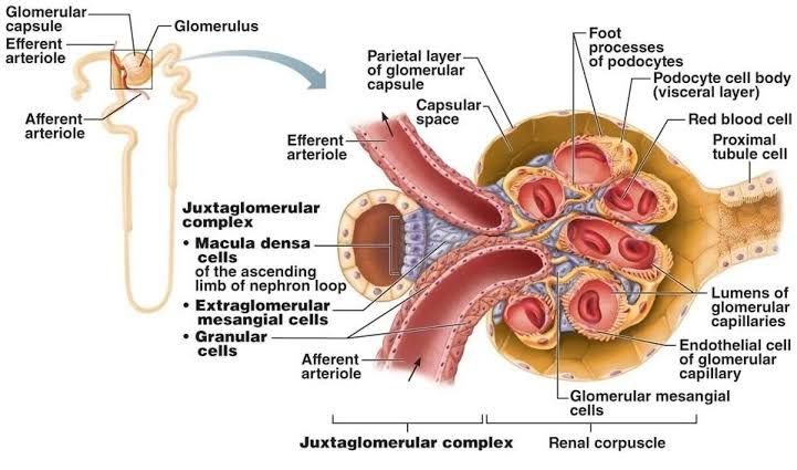
The juxtaglomerular apparatus (JGA) is a specialized anatomical structure located at the junction of the afferent and efferent arterioles with the initial part of the distal convoluted tubule of the same nephron. It serves as a critical sensing and signaling center involved in regulating blood pressure, blood volume, and electrolyte balance, primarily through the initiation of the renin-angiotensin system (RAS).
The JGA is composed of three main cell types:
- Macula Densa Cells: As discussed in Section 1, these are specialized epithelial cells located in the wall of the early distal convoluted tubule. They are positioned directly opposite the renal corpuscle’s vascular pole, in close contact with the afferent and efferent arterioles. Their primary known function is to sense the NaCl concentration and flow rate of the tubular fluid. They play a key role in tubuloglomerular feedback and influencing renin release.
- Granular Cells (Juxtaglomerular Cells): These are modified smooth muscle cells found predominantly in the wall of the afferent arteriole, just upstream from the glomerulus, but also sometimes in the efferent arteriole. They are the primary storage site and source of renin. Granular cells function as intrarenal baroreceptors, releasing renin in response to decreased stretch (indicating low blood pressure or RPP) in the afferent arteriole. They are also stimulated to release renin by signals from the macula densa (indicating low tubular NaCl/flow) and by sympathetic nerve activity (via β1-adrenergic receptors).
- Extraglomerular Mesangial Cells (Lacis Cells): Located in the triangular space between the macula densa and the afferent and efferent arterioles. Their exact function is not fully elucidated, but they are thought to be involved in communication between the macula densa and the granular cells, potentially mediating signals that regulate renin release and arteriolar tone.
Role of the JGA in Initiating the Renin-Angiotensin System (RAS):
The JGA is the primary control point for initiating the systemic renin-angiotensin system, a major hormonal cascade regulating blood pressure and fluid balance. Renin is the rate-limiting enzyme in this pathway.
-
- Renin Release: As mentioned, renin is released from granular cells in response to three main stimuli, all indicating potential hypovolemia or hypotension:
- Decreased stretch of the afferent arteriole (intrarenal baroreceptor).
- Decreased NaCl delivery to the macula densa (sensed via TGF mechanism).
- Increased sympathetic nerve activity to the kidney.
- The RAS Cascade: Once released into the bloodstream, renin acts on a protein called Angiotensinogen, which is continuously produced by the liver.
- Renin cleaves Angiotensinogen to form a relatively inactive decapeptide, Angiotensin I.
- Angiotensin I circulates in the blood and is converted to the highly active octapeptide, Angiotensin II, primarily by Angiotensin-Converting Enzyme (ACE), which is abundant in the endothelium of blood vessels, particularly in the lungs.
- Actions of Angiotensin II: Angiotensin II is a potent hormone with widespread effects that collectively work to increase blood pressure and restore fluid volume:
- Vasoconstriction: It is a powerful systemic vasoconstrictor, particularly of arterioles, which increases total peripheral resistance and raises mean arterial pressure. It also affects renal arterioles, often preferentially constricting the efferent arteriole at physiological concentrations, which helps maintain or even increase GFR when RPP is low.
- Aldosterone Release: Angiotensin II stimulates the adrenal cortex to release Aldosterone. Aldosterone acts on the principal cells of the collecting ducts and late distal tubules to increase sodium and water reabsorption and potassium secretion. This increases blood volume.
- ADH Release: It stimulates the release of Antidiuretic Hormone (ADH or Vasopressin) from the posterior pituitary gland. ADH increases water reabsorption in the collecting ducts, further increasing blood volume.
- Thirst Stimulation: Angiotensin II acts on the brain to stimulate thirst, promoting water intake.
- Sodium Reabsorption: It directly stimulates sodium reabsorption in the proximal tubules.
- By triggering the RAS, the JGA plays a vital role in long-term blood pressure regulation and maintaining extracellular fluid volume, linking renal filtration status to systemic hemodynamic and fluid balance responses.
- Renin Release: As mentioned, renin is released from granular cells in response to three main stimuli, all indicating potential hypovolemia or hypotension:
Glomerulotubular Balance
Glomerulotubular balance is a mechanism primarily operating in the proximal tubule that ensures that the rate of proximal tubular reabsorption is proportional to the rate of glomerular filtration. It means that a relatively constant fraction (percentage) of the filtered load of solutes and water is reabsorbed in the proximal tubule, irrespective of minor fluctuations in GFR. While renal autoregulation aims to keep GFR stable, small variations still occur. Glomerulotubular balance handles these variations by adjusting proximal reabsorption accordingly.
-
- The Concept: If GFR increases, the absolute amount (mass or volume) of fluid and solutes filtered into Bowman’s space – the filtered load – increases proportionally. Glomerulotubular balance dictates that the proximal tubule will then increase its reabsorption rate so that the fraction (around 65-70%) of the filtered load reabsorbed remains constant. For example, if GFR doubles, the filtered load doubles, and the proximal tubule approximately doubles its reabsorption rate. The absolute amount reabsorbed increases, but the percentage reabsorbed stays mostly the same.
- Mechanisms: The precise mechanisms are complex and involve several factors:
- Peritubular Capillary Starling Forces: Changes in GFR influence the hydrostatic and oncotic pressures in the peritubular capillaries that surround the proximal tubule.
- When GFR increases (often due to higher RPP or specific arteriolar resistance changes), the volume of plasma filtered out in the glomerulus increases. This leaves the blood flowing into the efferent arteriole and subsequently the peritubular capillaries with a lower hydrostatic pressure (Ppc) and a higher protein concentration, leading to a higher oncotic pressure (πpc).
- These altered Starling forces (low Ppc, high πpc) in the peritubular capillaries create a more favorable gradient for the reabsorption of fluid and solutes from the interstitial space (between the tubule and capillary) into the capillaries. This increased uptake drives increased net reabsorption from the tubular lumen across the proximal tubular cells.
- Filtered Load of Solutes: While Starling forces are considered a primary driver, the increased delivery of solutes like Na+ to the proximal tubule when GFR is high may also directly stimulate transport mechanisms in the proximal tubular cells, contributing to increased reabsorption.
- Peritubular Capillary Starling Forces: Changes in GFR influence the hydrostatic and oncotic pressures in the peritubular capillaries that surround the proximal tubule.
- Physiological Significance: Glomerulotubular balance is essential because it prevents large fluctuations in the delivery of fluid and solutes to the downstream segments of the nephron, particularly the loop of Henle, distal tubule, and collecting ducts. These downstream segments are responsible for the fine-tuning of electrolyte and water balance. Without glomerulotubular balance, even modest increases in GFR would lead to massive increases in fluid and solute delivery to the distal nephron, potentially overwhelming its capacity to reabsorb, resulting in excessive salt and water loss (e.g., diuresis and natriuresis). Conversely, decreases in GFR would lead to excessively low delivery. By keeping the delivery to the loop of Henle relatively constant proportionally, glomerulotubular balance allows the distal nephron to perform its precise regulatory roles more effectively and prevents detrimental imbalances in fluid and electrolyte excretion.
In conclusion, the kidney employs a sophisticated suite of integrated mechanisms – renal autoregulation (myogenic response and tubuloglomerular feedback), the juxtaglomerular apparatus acting as the control center for the renin-angiotensin system, and glomerulotubular balance – to precisely control the glomerular filtration rate and coordinate it with subsequent tubular processing. These mechanisms collectively ensure stable kidney function, contribute significantly to the regulation of systemic blood pressure and blood volume, and maintain the delicate balance of fluid and electrolytes crucial for overall physiological homeostasis.


