The digestive system is a complex network responsible for breaking down food into absorbable nutrients. A critical aspect of this process involves the secretion of various fluids containing enzymes, acids, mucus, and hormones into the lumen of the gastrointestinal tract. These secretions are precisely regulated to ensure efficient digestion and protection of the digestive lining. This guide will delve into the intricacies of gastric and intestinal secretions, exploring the cells responsible, the composition and function of the secreted fluids, the diverse control mechanisms at play, and the role of hormonal and other factors in modulating these vital processes.
The Cells of the Gastric Mucosa and Their Secretions
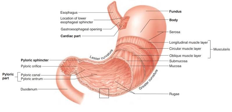
The stomach lining, or gastric mucosa, is a specialized epithelium containing different types of cells, each contributing unique components to gastric juice. These cells are primarily located within the gastric glands, which are invaginations of the surface epithelium.
- Parietal Cells (also known as Oxyntic Cells):
- Location: Fundus and body of the stomach glands.
- Secretions:
- Hydrochloric Acid (HCl): A highly acidic solution (pH 1.5-3.5).
- Intrinsic Factor: A glycoprotein essential for the absorption of Vitamin B12 in the ileum.
- Function: HCl provides the acidic environment necessary for activating pepsinogen, denaturing proteins, killing ingested microorganisms, and facilitating the absorption of certain minerals like iron and calcium. Intrinsic factor binds to dietary B12, forming a complex that is resistant to digestion and allows for its uptake.
- Chief Cells (also known as Peptic Cells):
- Location: Deeper parts of the gastric glands in the fundus and body.
- Secretions:
- Pepsinogen: An inactive zymogen (precursor enzyme).
- Gastric Lipase: An enzyme that contributes to the digestion of fats, though its activity is limited in the stomach’s acidic environment compared to pancreatic lipase.
- Function: Pepsinogen is converted to the active enzyme pepsin in the presence of HCl. Pepsin is a primary enzyme for the digestion of proteins into smaller polypeptides. Gastric lipase initiates the breakdown of triglycerides, particularly short- and medium-chain fatty acids.
- Mucous Neck Cells:
- Location: Neck region of the gastric glands, interspersed among parietal cells.
- Secretions: Soluble mucus and some bicarbonate.
- Function: This soluble mucus helps lubricate the gastric contents and protects the epithelium from mechanical damage. Along with surface mucous cells, they contribute to the bicarbonate buffer layer.
- Surface Mucous Cells:
- Location: Cover the entire surface epithelium of the stomach.
- Secretions: Thick, alkaline mucus and bicarbonate.
- Function: These cells form a protective gel-like layer on the gastric surface. Bicarbonate trapped within this mucus layer creates a pH gradient, with a near-neutral pH zone adjacent to the epithelium, buffering against the harsh acidity of the gastric lumen and preventing autodigestion.
- Enteroendocrine Cells: These are scattered throughout the gastric mucosa and secrete hormones into the bloodstream.
- G Cells: Located primarily in the pyloric antrum. Secrete Gastrin, a hormone that stimulates HCl secretion (indirectly by stimulating ECL cells and directly on parietal cells) and pepsinogen secretion.
- D Cells: Located in the antrum and body. Secrete Somatostatin, a hormone that inhibits gastric secretion (HCl, gastrin, histamine) and gastric motility.
- ECL Cells (Enterochromaffin-like Cells): Located in the body and fundus. Stimulated by gastrin and acetylcholine, they secrete Histamine, a potent paracrine mediator that significantly stimulates HCl secretion by binding to H2 receptors on parietal cells.
Deconstructing Gastric Juice: Components and Functions
Gastric juice is the collective secretion of the gastric glands, along with surface mucus. Its key components and their functions are:
- Hydrochloric Acid (HCl):
- Origin: Parietal cells.
- Function: Creates acidic environment (pH 1.5-3.5) essential for:
- Activation of pepsinogen to pepsin.
- Denaturation of proteins, exposing peptide bonds to enzymatic action.
- Killing most bacteria and other pathogens ingested with food.
- Facilitating absorption of iron (reduces ferric to ferrous form) and calcium.
- Contributing to the regulation of gastric emptying.
- Pepsinogen (converted to Pepsin):
- Origin: Chief cells.
- Function: Pepsin is an endopeptidase that begins protein digestion by hydrolyzing peptide bonds, breaking down large proteins into smaller polypeptides and some amino acids. It functions optimally in the acidic environment created by HCl.
- Mucus:
- Origin: Surface mucous cells and mucous neck cells.
- Function: Forms a protective layer covering the gastric epithelium. This layer, thickened by bicarbonate trapping, shields the underlying cells from mechanical damage, the corrosive effects of HCl, and the proteolytic action of pepsin.
- Intrinsic Factor:
- Origin: Parietal cells.
- Function: Binds to Vitamin B12 (cobalamin) in the stomach. This complex protects B12 from degradation and is necessary for its absorption in the terminal ileum via specific receptors. Deficiency in intrinsic factor leads to pernicious anemia.
- Water:
- Origin: Primarily secreted by parietal cells and surface mucous cells.
- Function: Provides the aqueous medium for enzymatic reactions and the suspension of gastric contents.
- Gastric Lipase:
- Origin: Chief cells.
- Function: Initiates the hydrolysis of some dietary triglycerides, especially those containing short- and medium-chain fatty acids, although its contribution is minor compared to pancreatic lipase.
Hormonal and Other Factors Influencing Gastric Secretion
Gastric secretion is not continuous; it varies based on the presence and nature of food in the stomach and the anticipation of eating. Several factors, particularly hormones and paracrine substances, play crucial roles in modulating the activity of gastric cells.
- Stimulatory Factors:
- Gastrin: Released from G cells in response to peptides, amino acids, distension of the stomach, and vagal stimulation (Gastrin-Releasing Peptide -GRP- from vagal endings). Gastrin stimulates parietal cells (directly and indirectly via ECL cells) and chief cells, increasing HCl and pepsinogen secretion.
- Histamine: Released from ECL cells, primarily stimulated by gastrin and acetylcholine. Histamine is a potent stimulator of HCl secretion by binding to H2 receptors on parietal cells. Gastrin and acetylcholine potentiate the effect of histamine, demonstrating synergy.
- Acetylcholine (ACh): Released from parasympathetic (vagal) nerve endings and neurons of the enteric nervous system. ACh stimulates parietal cells (via muscarinic receptors), chief cells, ECL cells, and G cells, increasing HCl, pepsinogen, histamine, and gastrin release, respectively.
- Peptides and Amino Acids: Directly stimulate G cells to release gastrin.
- Stomach Distension: Activates mechanoreceptors, triggering neural reflexes (local and vagovagal) that stimulate gastric secretion.
- Inhibitory Factors:
- Somatostatin: Released from D cells in the stomach (antrum and body) and duodenum. Stimulated by increased luminal acidity (low pH). Somatostatin acts locally (paracrine) to inhibit the release of gastrin, histamine, and acetylcholine, thereby reducing HCl and pepsinogen secretion. It acts as a critical negative feedback mechanism.
- Low pH in the Antrum: Directly inhibits G cell activity and thus gastrin release, independent of somatostatin.
- Low pH, Fats, Carbohydrates, and Hypertonicity in the Duodenum: When acidic chyme enters the duodenum, it triggers inhibitory mechanisms that slow gastric emptying and secretion. These include:
- Enterogastric Reflex: Neural reflex mediated by the enteric nervous system and extrinsic nerves.
- Enterogastrones: Hormones released from the duodenum and jejunum in response to duodenal contents. Key enterogastrones include:
- Secretin: Released in response to acidic chyme. Inhibits gastric acid secretion and stimulates pancreatic bicarbonate secretion.
- Cholecystokinin (CCK): Released in response to fats and proteins. Primarily stimulates pancreatic enzyme secretion and gallbladder contraction, but also inhibits gastric emptying and acid secretion.
- Gastric Inhibitory Peptide (GIP): Released in response to glucose and fat. Inhibits gastric acid secretion and motility; also stimulates insulin release (hence its alternative name, Glucose-dependent Insulinotropic Peptide).
Control Mechanisms of Gastric Secretion: The Three Phases
Gastric secretion is regulated by a complex interplay of neural, hormonal, and local chemical signals, traditionally divided into three overlapping phases based on the location of the stimulus:
- Cephalic Phase:
- Stimulus: Sight, smell, taste, thought, or sound of food. Originate in the brain.
- Mechanism: Primarily neural. Signals from the cerebral cortex, hypothalamus, and medulla oblongata are transmitted via the vagus nerve (parasympathetic innervation) to the stomach.
- Effects:
- Vagus nerve (via ACh) stimulates parietal cells (minor direct effect), chief cells, ECL cells, and G cells (via GRP).
- Results in increased HCl, pepsinogen, histamine, and gastrin secretion.
- Contribution: Accounts for about 20-30% of the total acid secretion response to a meal. Prepares the stomach for incoming food.
- Gastric Phase:
- Stimulus: Food entering the stomach. Stimuli within the stomach itself.
- Mechanism: Neural (local and vagovagal reflexes), hormonal, and chemical.
- Distension: Stretching of the stomach wall activates mechanoreceptors. This triggers short (enteric nervous system) and long (vago-vagal) reflexes, leading to increased ACh release and stimulation of gastric secretion.
- Peptides and Amino Acids: Breakdown products of protein digestion directly stimulate G cells in the antrum to release gastrin.
- Low Acidity: As food enters and buffers stomach acid, the pH rises. This buffering removes the inhibitory effect of low pH on G cells, allowing gastrin release.
- Effects: Significant increase in HCl and pepsinogen secretion. Gastrin is a major player in this phase, stimulating both parietal and chief cells, and amplifying the neural input.
- Contribution: The most significant phase, accounting for 50-60% of total acid secretion.
- Intestinal Phase:
- Stimulus: Chyme entering the duodenum. Primarily the presence of acid, fats, carbohydrates, and osmolarity changes in the duodenal lumen.
- Mechanism: Neural (enterogastric reflex) and hormonal (release of enterogastrones from duodenal/jejunal endocrine cells).
- Effects: Primarily inhibitory on gastric secretion and motility.
- Low pH in the duodenum stimulates Secretin release, which inhibits gastric acid secretion.
- Fats and protein digestion products stimulate CCK release, which inhibits gastric emptying and acid secretion.
- Glucose and fats stimulate GIP release, which inhibits gastric acid secretion and motility.
- The enterogastric reflex also contributes to the inhibition of gastric function.
- Contribution: Initially may have a brief stimulatory component (e.g., duodenal gastrin release), but quickly becomes predominantly inhibitory, accounting for the remaining 10-20% of the response and importantly, regulating the rate at which chyme enters the small intestine.
Intestinal Secretions and Their Control
As chyme moves from the stomach into the small intestine, the process of digestion and absorption continues, supported by intestinal secretions.
- Components of Intestinal Secretion (Succus Entericus):
- Water and Electrolytes: Large volumes of fluid (up to 1.5-2 liters per day) containing electrolytes like Na+, K+, Cl-, and importantly, bicarbonate (especially from Brunner’s glands in the duodenum and crypts of Lieberkühn throughout the small intestine).
- Mucus: Secreted by goblet cells scattered throughout the epithelium. Protects the intestinal lining from mechanical damage and chemical irritation.
- Brush Border Enzymes: While often listed as components, most digestive enzymes in the small intestine (like sucrase, lactase, maltase, peptidases, alkaline phosphatase) are bound to the brush border (microvilli) of the enterocytes rather than secreted freely into the lumen in significant quantities. Their activity occurs at the surface of the epithelial cells as nutrients are absorbed.
- Hormones: Enteroendocrine cells scattered throughout the intestinal epithelium secrete hormones (Secretin, CCK, GIP, VIP, Somatostatin, Motilin, etc.) into the bloodstream. These hormones regulate various digestive functions including motility, secretions from the pancreas and liver, and gastric activity (as discussed in Step 3). They are secreted by the intestine but primarily act on other organs or systems, not components of the luminal fluid itself, except perhaps indirectly by influencing water/electrolyte secretion.
- Control of Intestinal Secretion:
- Mechanical Stimulation: Distension of the intestinal wall by chyme activates local reflexes in the enteric nervous system, stimulating secretion of water, electrolytes, and mucus.
- Chemical Stimulation: The presence of acidic chyme, breakdown products of digestion (especially fats), and hypertonic solutions in the lumen stimulates both local neural reflexes and the release of hormones.
- Acidity: Stimulates Secretin release, which promotes bicarbonate secretion, neutralizing the chyme.
- Fats: Stimulate CCK release, which influences overall digestion but also has some inhibitory effects on intestinal motility in areas.
- Vasoactive Intestinal Peptide (VIP): A neuropeptide released from enteric neurons, strongly stimulates intestinal water and electrolyte secretion.
- Neural Control: Local reflexes within the enteric nervous system are the primary neural regulators. Parasympathetic (vagal) stimulation can enhance secretion, while sympathetic stimulation generally inhibits it.
- Hormonal Control: While hormones like Secretin and VIP directly influence water and electrolyte secretion, other hormones secreted by the intestine like CCK and GIP indirectly affect the overall intestinal environment by regulating upstream processes (gastric emptying, pancreatic/biliary secretion). Somatostatin acts locally to inhibit intestinal secretion.
Conclusion
The coordinated secretion of gastric and intestinal juices is fundamental to effective digestion. From the highly acidic, enzyme-rich environment of the stomach, carefully produced by specialized cells and tightly controlled by neural and hormonal inputs, to the bicarbonate-rich, protective secretions of the intestine facilitating final breakdown and absorption, these processes are precisely regulated. Understanding the diverse cellular origins, chemical compositions, functional roles, and intricate control mechanisms involving neural reflexes, local stimuli, and a symphony of hormones provides critical insight into the complex physiology of the gastrointestinal tract and the maintenance of digestive health.


