Understanding Alloimmunity in Transplantation
The success of organ transplantation hinges significantly on controlling the recipient’s immune response to the transplanted tissue (allograft). While immunosuppressive therapies are crucial, a thorough understanding of how the immune system recognizes and reacts to foreign antigens from the donor is paramount for clinicians and researchers. This guide provides a structured explanation of key concepts central to alloimmunity: antigen presentation pathways, the nature of allosensitization, the role of inflammation, and a critical marker of graft rejection.
The Foundation – Antigen Presentation
At the heart of adaptive immunity, including the response to transplanted organs, is the process of antigen presentation. This is how immune cells, particularly T lymphocytes (T cells), recognize foreign substances (antigens). Antigens are typically protein fragments (peptides) displayed on the surface of cells by specialized molecules called Major Histocompatibility Complex (MHC), known as Human Leukocyte Antigens (HLA) in humans. There are two main classes of MHC/HLA molecules:
- MHC Class I: Found on almost all nucleated cells. They primarily present peptides derived from proteins synthesized within the cell (e.g., viral proteins, tumor proteins, or normal cellular proteins). They are recognized by CD8+ cytotoxic T cells.
- MHC Class II: Primarily found only on professional Antigen-Presenting Cells (APCs), such as dendritic cells, macrophages, and B cells. They present peptides derived from proteins taken up by the cell from its external environment (e.g., bacterial proteins, soluble antigens). They are recognized by CD4+ helper T cells.
In transplantation, the significant difference lies in the fact that the donor graft contains donor MHC/HLA molecules, which are recognized as foreign by the recipient’s immune system. The way these foreign donor antigens are presented orchestrates the recipient’s response.
The Direct Pathway of Alloantigen Presentation
The direct pathway is the primary mechanism for the initial, rapid immune response against an allograft.
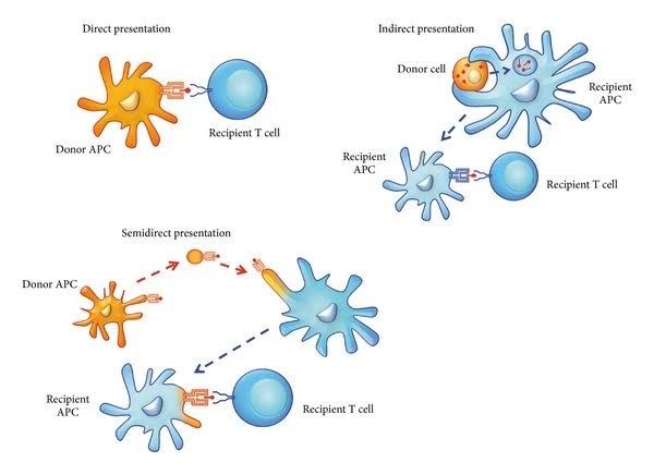
- Mechanism: In this pathway, recipient T cells directly recognize intact donor MHC molecules displayed on the surface of donor-derived APCs that have migrated from the graft into the recipient’s secondary lymphoid organs (like lymph nodes or spleen). These donor APCs (often passenger lymphocytes or dendritic cells within the transplanted organ) are highly effective at stimulating recipient T cells because they present the donor’s own MHC molecules.
- Antigen: The ‘antigen’ here can be considered the entire donor MHC molecule itself, potentially complexed with donor self-peptides. Recipient T cells recognize the donor MHC structure as foreign.
- Key Players: Donor APCs (within the graft), recipient CD4+ T cells, and recipient CD8+ T cells.
- Outcome: Direct recognition bypasses the need for recipient processing of donor antigens and is highly efficient at activating a large number of recipient T cells (a phenomenon known as “alloreactivity”). A significant percentage (1-10%) of a recipient’s T cell repertoire can potentially react directly against a foreign MHC molecule, compared to the much smaller fraction (<0.001%) that would recognize a foreign peptide presented by self MHC. This leads to a potent and rapid cellular response, primarily mediated by activated recipient T cells targeting the graft. CD8+ T cells can directly kill graft cells expressing donor MHC Class I, while CD4+ T cells provide help for the activation of CD8+ T cells and B cells.
The Indirect Pathway of Alloantigen Presentation
The indirect pathway contributes to the later, more sustained chronic alloimmune response, including the development of donor-specific antibodies (DSA).
- Mechanism: In this pathway, recipient professional APCs (dendritic cells, macrophages) internalize and process donor graft antigens. These antigens can be shed from dying graft cells, released from matrix breakdown, or taken up from circulating donor material. The recipient APCs then present peptides derived from donor proteins (including fragments of donor MHC molecules) on their own recipient MHC Class II molecules.
- Antigen: Peptides originating from donor proteins (including donor MHC/HLA proteins) presented on recipient MHC Class II molecules.
- Key Players: Recipient professional APCs, recipient CD4+ T cells.
- Outcome: Recipient CD4+ T cells recognize these donor peptides presented by self-MHC. This interaction is crucial for activating helper T cells, which in turn support the activation of cytotoxic T cells and, importantly, provide help to B cells. B cell activation leads to the differentiation of plasma cells that produce antibodies against donor antigens, including donor HLA molecules (anti-HLA antibodies). The indirect pathway is slower to develop than the direct pathway but is essential for generating immunological memory and mediating chronic rejection, often involving antibody-mediated mechanisms. It can also contribute to cellular responses via helper T cell support.
Distinguishing Between Direct and Indirect Pathways
| Feature | Direct Pathway | Indirect Pathway |
|---|---|---|
| Antigen Presenting Cell (APC) | Donor APCs (from the graft) | Recipient Professional APCs |
| MHC Molecule Used | Donor MHC Class I and Class II | Recipient MHC Class II (predominantly) |
| Antigen Presented | Intact Donor MHC (potentially with donor self-peptides) | Peptides derived from donor proteins (including donor MHC) |
| Recipient T Cell Recognition | Recipient T cell receptor recognizes the entire donor MHC molecule | Recipient T cell receptor recognizes a donor peptide presented by recipient MHC |
| Speed of Activation | Rapid, initial strong response | Slower, develops over time |
| Predominant T Cell Subset | Activates both CD4+ and CD8+ T cells efficiently | Primarily activates CD4+ T helper cells, which support B cell and CD8+ responses |
| Role in Rejection | Primarily initiates acute cellular rejection | Contributes to both cellular and humoral (antibody-mediated) rejection; essential for DSA formation and chronic rejection |
| Immunological Memory | Can contribute, but indirect pathway is key for long-term humoral memory | Crucial for generating long-term T cell and B cell memory and antibody responses (DSA) |
In essence, the direct pathway is like a recipient T cell recognizing the face (donor MHC) of the foreign donor APC, triggering an immediate alarm. The indirect pathway is like recipient APCs processing parts of the foreign tissue and presenting small fragments (donor peptides) on their own faces (recipient MHC), leading to a more nuanced, antibody-focused, and long-term response.
An overview of Allosensitization
Allosensitization is the process by which a recipient’s immune system becomes primed or sensitized to alloantigens (antigens from another individual of the same species). In the context of transplantation, this specifically refers to developing an immune response (cellular and/or humoral) against donor alloantigens, most notably the donor’s HLA molecules.
Prior exposure to alloantigens can lead to allosensitization. Common ways this occurs include:
- Previous blood transfusions
- Previous transplants or tissue grafts
- Pregnancy (exposure to paternal antigens from the fetus)
Allosensitization is clinically measured by the presence of donor-specific antibodies (DSA) in the recipient’s serum, often detected using solid-phase assays that test for antibodies against specific HLA alleles. A highly sensitized patient (high panel reactive antibody – PRA, or presence of specific DSA) is at increased risk of immediate or accelerated rejection due to pre-existing immunity.
Distinguishing Allosensitization from Other Immune Responses
While the immune system uses similar mechanisms (T cells, B cells, antibodies) for various responses, the target antigen defines the type of reaction:
- Response to Foreign Pathogens (e.g., Bacteria, Viruses): The target antigens are microbial components (PAMPs) or proteins expressed by pathogens. The goal is pathogen clearance.
- Response to Tumors: The target antigens are mutated self-proteins or aberrantly expressed self-proteins (neoantigens, tumor-associated antigens). The goal is tumor cell destruction.
- Autoimmunity: The target antigens are normal self-proteins or tissues. The response is a breakdown of self-tolerance, leading to attack on one’s own body.
- Allergy: The target antigens are typically harmless environmental substances (allergens). The response involves IgE antibodies and mast cell degranulation.
- Alloimmunity (Allosensitization/Rejection): The target antigens are foreign antigens from a genetically different individual of the same species, predominantly MHC/HLA molecules and minor histocompatibility antigens. The goal (from the immune system’s perspective) is to reject the foreign tissue/organ, regardless of whether it is beneficial to the host.
Allosensitization is unique as it involves a response directed specifically against non-self antigens within the same species, resulting from prior exposure and leading to a memory response that can threaten a subsequent allograft bearing those antigens. Unlike autoimmunity which targets self, or responses to pathogens/tumors which target truly foreign/mutated entities for clearance, alloimmunity targets healthy, functional tissue from another human.
Inflammatory Factors Contributing to the Efficient Immune Response to Alloantigen in a Transplant Setting
The transplantation process itself is inherently pro-inflammatory, significantly amplifying the alloimmune response. Several factors contribute to this inflammatory milieu:
- Ischemia-Reperfusion Injury (IRI): The period when the organ is deprived of blood flow (ischemia during transport and surgery) followed by restoration of flow (reperfusion) causes significant cellular damage. This injury releases intracellular contents, including Damage-Associated Molecular Patterns (DAMPs).
- Damage-Associated Molecular Patterns (DAMPs): Molecules released from injured or dying graft cells (e.g., HMGB1, heat shock proteins, ATP, uric acid) act as “danger signals.” They activate innate immune receptors (like TLRs) on recipient immune cells and resident cells in the graft, triggering a robust inflammatory response.
- Surgical Trauma: The physical process of surgery causes tissue injury and inflammation in the recipient.
- Release of Pro-inflammatory Cytokines and Chemokines: Activated resident cells in the graft (endothelial cells, macrophages) and recruited recipient immune cells release a cascade of inflammatory mediators (e.g., TNF-alpha, IL-1 beta, IL-6, IL-12, IFNs, chemokines like CXCL10, CCL2). These attract, activate, and mature recipient APCs and T cells.
- Activation and Maturation of Recipient APCs: Inflammatory signals (DAMPs, cytokines) cause recipient APCs to mature. Mature APCs upregulate co-stimulatory molecules (like CD80, CD86) and MHC molecules, making them highly effective at presenting donor antigens (via the indirect pathway) and activating recipient T cells. They also migrate efficiently to draining lymph nodes.
- Presence of Donor APCs: As discussed in the direct pathway, donor APCs migrate from the graft and initiate a strong early response.
- Complement Activation: Graft injury and bound antibodies can activate the complement system, generating inflammatory mediators (anaphylatoxins like C3a, C5a) that increase vascular permeability and recruit immune cells.
These inflammatory signals create a highly activating environment for the recipient’s immune system, lowering the threshold for T cell activation and enhancing the efficiency of both direct and indirect antigen presentation pathways, thereby fueling the alloimmune response leading to potential rejection.
The Significance of C4d Positivity on an Allograft Biopsy
An allograft biopsy is a critical tool for diagnosing rejection and monitoring graft health. Finding C4d deposits on the biopsy tissue is a significant clinical finding.
- What is C4d? C4d is a stable cleavage product of the complement protein C4. It is generated when the classical or lectin pathways of the complement system are activated. Specifically, C4 is cleaved into C4a and C4b, and C4b can then be cleaved further into C4d and C4c. Unlike other complement fragments, C4d covalently binds to tissue surfaces and persists for extended periods (days to weeks), making it a reliable marker of prior or ongoing complement activation at the site of deposition.
- Mechanism in Transplantation: In the context of allograft rejection, C4d deposition most commonly occurs when antibodies, particularly donor-specific antibodies (DSA) directed against graft endothelium (e.g., anti-HLA antibodies), bind to targets on the graft vasculature. The binding of DSA (most commonly IgG antibodies like IgG1, IgG2, IgG3, or IgM) can activate the classical complement pathway via the C1 complex (C1q, C1r, C1s). This activation cascade involves the cleavage of C4 into C4a and C4b, followed by the cleavage of C2 into C2a and C2b, forming the C3 convertase (C4bC2a). This leads to C3 activation, and downstream effects which can include cell lysis, inflammation, and opsonization. However, even if the full cascade isn’t completed, the C4b generated can bind covalently to tissue, leading to the stable C4d fragment upon further cleavage of C4b.
- Significance: C4d positivity on an allograft biopsy is considered a histopathological marker strongly indicative of antibody-mediated rejection (AMR). Its presence signifies that complement, specifically via pathways involving C4 activation (most often initiated by antibody binding), has been activated within the graft tissue. Along with other histological features of injury (e.g., microvascular inflammation) and the presence of circulating donor-specific antibodies (DSA), C4d positivity is a diagnostic criterion for AMR according to the Banff classification system, which is the standard for evaluating transplant rejection. While not the sole criterion, C4d positivity is a crucial piece of evidence supporting a diagnosis of AMR, which often requires different treatment strategies compared to T cell-mediated rejection.
In summary, C4d serves as a “footprint” left behind by complement activation, primarily triggered by antibodies binding to the graft vasculature, making it a vital indicator of humoral injury and antibody-mediated rejection in transplanted organs.


