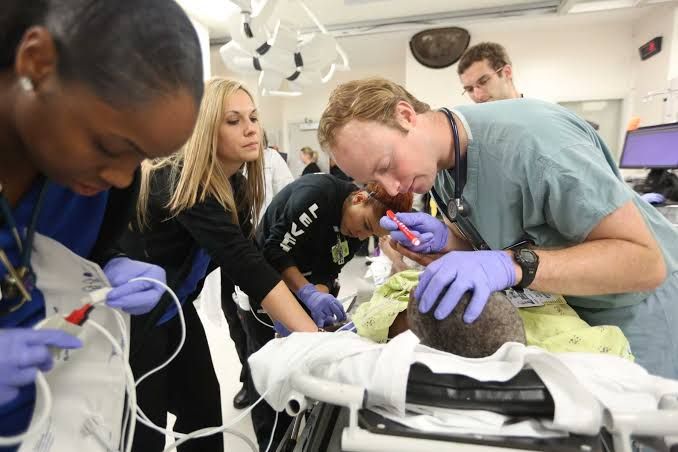
Effective trauma care relies on a systematic, prioritized approach aimed at rapidly identifying and managing life-threatening conditions to optimize patient outcomes. The principles outlined below are foundational to this process, drawing heavily from established frameworks such as Advanced Trauma Life Support (ATLS) and Primary Trauma Care (PTC).
Principles of Advanced Trauma Life Support (ATLS) and Primary Trauma Care (PTC)
These systems provide a standardized approach to trauma patient management, emphasizing a rapid, systematic assessment and resuscitation sequence. The core philosophy is to treat the greatest threat to life first.
-
- Systematic Approach: A structured sequence ensures that critical issues are not missed. This sequence includes the Primary Survey, Resuscitation, Secondary Survey, and Definitive Care considerations.
- Primary Survey (ABCDE): This is the most critical initial step, focusing on identifying and managing immediate life threats.
- A – Airway Maintenance with Cervical Spine Protection: Ensure a patent airway. Look for signs of obstruction (stridor, hoarseness, inability to speak). If the airway is compromised, establish one using maneuvers (jaw thrust/chin lift – with c-spine control), suction, or definitive methods like intubation. Always assume cervical spine injury in blunt multi-trauma until cleared.
- B – Breathing and Ventilation: Assess breathing adequacy. Look for chest symmetry, respiratory rate, effort, and listen to breath sounds. Identify and manage immediate threats like tension pneumothorax (needle decompression), massive hemothorax (chest tube), or flail chest with paradoxical movement (ventilation support). Oxygen administration is standard.
- C – Circulation with Hemorrhage Control: Assess circulatory status (pulse, skin color, capillary refill, blood pressure). Identify and control external hemorrhage promptly through direct pressure. Establish intravenous access (two large-bore IVs) and initiate fluid resuscitation (crystalloids, potentially blood products early in massive hemorrhage). Look for signs of internal bleeding.
- D – Disability (Neurologic Status): Perform a rapid neurologic assessment. Use the AVPU scale (Alert, Verbal, Pain, Unresponsive) or Glasgow Coma Scale (GCS). Assess pupillary size and reaction. This provides a baseline and can indicate head injury or other neurological compromise.
- E – Exposure and Environment Control: Completely expose the patient to perform a thorough physical examination, looking for injuries. Prevent hypothermia by covering the patient with warm blankets and using warmed fluids.
- Resuscitation Adjuncts: These are tools and tests used to support the primary survey and guide resuscitation (e.g., oxygen saturation monitoring, capnography, ECG, Foley catheter, nasogastric tube, focused assessment with sonography for trauma [FAST], chest X-ray, pelvic X-ray).
- Secondary Survey: Once the primary survey is complete, life threats are addressed, and the patient is relatively stable, a head-to-toe examination is performed, along with a comprehensive history (AMPLE: Allergies, Medications, Past medical history/Pregnancy, Last meal, Events leading to injury). This phase aims to identify all other injuries.
- Ongoing Assessment: The patient’s status must be continually monitored as conditions can change rapidly. Reassess the ABCDEs frequently.
- Definitive Care: This involves organizing the patient’s ongoing management, which might include transfer to a higher level trauma center, surgical intervention, or admission to an appropriate ward or intensive care unit.
Triage and Identification/Management of Life-Threatening Injuries
Triage is the process of sorting patients based on the severity of their injuries and their need for immediate medical attention. In the trauma setting, particularly in mass casualty incidents or overloaded facilities, effective triage is crucial for allocating limited resources to maximize patient survival.
-
- Purpose of Triage: To identify patients with immediate life threats who require urgent stabilization and intervention versus those with less severe injuries who can wait.
- Triage Principles: Systems often use simple algorithms (e.g., based on respiratory rate, perfusion, mental status) to categorize patients into priority groups (e.g., Immediate, Delayed, Minor, Expectant).
- Identification of Life-Threatening Injuries: The primary survey is the cornerstone of identifying immediate life threats. Key conditions requiring urgent management include:
- Airway Obstruction: Managed with maneuvers, suction, or definitive airway control.
- Severe External Hemorrhage: Controlled by direct pressure, elevation, and potentially tourniquets in extremity injuries.
- Respiratory Distress/Failure: May indicate tension pneumothorax (requires needle decompression followed by chest tube), massive hemothorax (requires chest tube and fluid/blood resuscitation), or other compromised ventilation. Initial management focuses on support and decompression/drainage.
- Signs of Shock: Rapid pulse, low blood pressure, poor perfusion. Assume hypovolemic shock from blood loss until proven otherwise. Initial management involves controlling hemorrhage and aggressive fluid/blood resuscitation.
- Altered Mental Status: Can indicate head injury or hypoperfusion. Requires airway protection and investigation into cause.
- Urgent Management: Life-threatening conditions identified in the primary survey are managed immediately as they are found, before proceeding to the next step of the primary survey or the secondary survey. This “treat as you go” approach is fundamental.
Differentiating Between the Mechanism of Blunt and Penetrating Trauma
Understanding the mechanism of injury (MOI) is vital in trauma assessment as it helps predict potential injury patterns and maintain a high index of suspicion for specific injuries.
-
- Blunt Trauma: Occurs when energy is transferred to the body without penetrating the skin. Common causes include motor vehicle crashes (MVCs), falls from height, pedestrian struck by vehicle, and direct blows.
- Injury Pattern: Injuries from blunt trauma are often widespread and may not have obvious external signs. Energy transfer can cause:
- Compression Injuries: Organs or tissues crushed between external force and bony structures.
- Shear Injuries: Tissues or organs tearing due to differential movement (e.g., aortic injury in rapid deceleration MVCs).
- Deceleration/Acceleration Injuries: Organs moving within body cavities, impacting bony structures or ligaments.
- Blast Injuries: Complex blunt and penetrating effects from explosive events.
- Assessment Implications: Requires a high index of suspicion for internal injuries despite minimal external findings. Thorough physical examination and imaging (X-rays, CT scans) are often necessary.
- Injury Pattern: Injuries from blunt trauma are often widespread and may not have obvious external signs. Energy transfer can cause:
- Penetrating Trauma: Occurs when an object pierces the skin and soft tissues, creating a wound. Common causes include stabbings (knives, ice picks) and gunshot wounds (GSWs).
- Injury Pattern: Injuries typically follow the path of the penetrating object.
- Stab Wounds: Injury path is relatively linear, but internal damage depth and angle can be misleading. Organ injury depends on location and depth.
- Gunshot Wounds (GSWs): More complex. Energy transfer causes direct tissue damage along the bullet path, but also creates a “temporary cavity” around the path due to kinetic energy dissipation, causing damage to surrounding tissue. Bullet fragmentation and ricochet within the body add complexity. High-velocity GSWs cause significantly more damage than low-velocity ones.
- Assessment Implications: Requires careful assessment of entry and exit wounds (if present) and predicting the potential path of the object to identify damaged organs or structures. Imaging (X-rays to locate foreign bodies, CT scans) helps delineate the extent of injury.
- Injury Pattern: Injuries typically follow the path of the penetrating object.
- Blunt Trauma: Occurs when energy is transferred to the body without penetrating the skin. Common causes include motor vehicle crashes (MVCs), falls from height, pedestrian struck by vehicle, and direct blows.
Compartment Syndrome and Its Management
Compartment syndrome is a limb-threatening and potentially life-threatening condition caused by increased pressure within a closed fascial compartment, leading to reduced tissue perfusion and subsequent ischemia and potential necrosis.
-
- Pathophysiology: Trauma (fractures, crush injuries, severe contusions) or other causes (burns, constrictive dressings, reperfusion injury) lead to bleeding or swelling within a fascial compartment. The rigid fascia prevents outward expansion, causing pressure to rise. When compartment pressure exceeds capillary perfusion pressure, muscle and nerve tissue become ischemic. Continued ischemia leads to cellular damage, capillary leak, and more swelling, creating a vicious cycle.
- Common Locations: Most often affects the compartments of the lower leg and forearm, but can occur anywhere enclosed by fascia.
- Clinical Features (The 5 Ps):
- Pain: Often described as severe, deep, and out of proportion to the apparent injury. Characteristically worsens with passive stretching of the muscles within the affected compartment. This is often the earliest and most reliable sign.
- Paresthesia: Numbness or tingling in the distribution of nerves running through the compartment.
- Pallor: Skin may appear pale, although this is less reliable as a sign.
- Paralysis: Weakness or inability to move the muscles within the compartment (a later and concerning sign).
- Pulselessness: Absence of a palpable pulse distally (a very late and ominous sign indicating significant arterial compromise, often preceding irreversible damage).
- Diagnosis: Primarily clinical, based on the signs and symptoms. Compartment pressure measurement can be used to confirm the diagnosis, especially in unreliable patients (e.g., altered mental status, intubated) or when clinical signs are equivocal. Pressures significantly elevated above diastolic blood pressure or an absolute pressure >30-40 mmHg are generally considered diagnostic.
- Management:
- Immediate Measures: Remove any constrictive dressings or casts. Elevate the limb to heart level (elevating significantly above heart level can reduce arterial inflow and worsen ischemia). Administer analgesia.
- Definitive Management: The definitive treatment for established compartment syndrome is emergency fasciotomy. This involves surgical incision through the fascia to release the pressure, restore blood flow, and prevent irreversible nerve and muscle damage.
- Timing is Critical: Delayed diagnosis and fasciotomy can lead to permanent disability (contractures, nerve damage) or even limb loss. Muscle necrosis begins after a few hours of ischemia.
Management of Abdominal Trauma (Liver, Spleen, Intestines)
Abdominal trauma is a major cause of morbidity and mortality. Injuries can cause significant internal bleeding (solid organs, major vessels) and/or peritonitis (hollow organ perforation). Assessment is challenging as signs can be subtle or delayed.
-
- Assessment: Suspect abdominal trauma in any significant blunt or penetrating injury to the torso. Assessment includes physical examination (tenderness, guarding, rebound, distension, seat belt sign, external wounds), and diagnostic adjuncts like FAST scan (rapidly assesses for free fluid/blood in the abdomen/pelvis), Diagnostic Peritoneal Lavage (DPL – less common now, but useful in unstable patients), and CT scan (most informative for identifying specific organ injuries and their grade in stable patients).
- General Management Principles:
- Resuscitation: Aggressive fluid and blood product resuscitation is critical for hypovolemic shock due to internal bleeding.
- Identify Source: Determine if bleeding is occurring and if there is contamination from hollow organ injury.
- Decision for Intervention: Decide between operative (surgery) and non-operative management based on patient stability, type and severity of injury, and findings on imaging. Unstable patients with suspected abdominal bleeding or peritonitis typically require immediate surgery.
- Specific Organ Management Principles:
- Liver: The most commonly injured solid organ in blunt trauma. Injuries range from minor lacerations to severe parenchymal disruption and vascular injury.
- Management: Many blunt liver injuries in hemodynamically stable patients can be managed non-operatively with close monitoring (serial exams, hemoglobin) and bed rest. Interventional radiology (angioembolization) is useful for controlling bleeding in some cases. Surgical management (packing, repair, or less commonly, resection) is required for hemodynamically unstable patients with ongoing bleeding or very high-grade injuries not controlled by embolization, or with associated injuries requiring surgery.
- Spleen: The most commonly injured organ in blunt trauma overall. Injuries range from contusions to shattered spleen.
- Management: Non-operative management (NOM) in hemodynamically stable patients has become the standard of care for most blunt splenic injuries, especially in lower grades. NOM involves monitoring in a trauma setting, serial blood counts, and potentially angioembolization to control bleeding. Splenectomy (removal of the spleen) or splenic repair is reserved for hemodynamically unstable patients who don’t respond to resuscitation, have ongoing bleeding despite embolization, or have high-grade injuries with associated instability. Patients who undergo splenectomy require post-splenectomy vaccinations due to increased risk of overwhelming infection.
- Intestines (Small and Large Bowel): More common in penetrating trauma, but blunt trauma (especially deceleration or compression against the spine/pelvis, like from seatbelts) can cause rupture or contusion. High risk of contamination leading to peritonitis.
- Management: Injuries typically require surgical intervention. Simple perforations can often be repaired. More extensive damage or multiple perforations may require resection of the damaged segment with primary anastomosis (reconnecting the ends) or creation of a stoma (ostomy) to divert fecal flow, particularly in the colon, in unstable patients, or with significant contamination. Aggressive irrigation and broad-spectrum antibiotics are crucial to manage bacterial contamination.
- Liver: The most commonly injured solid organ in blunt trauma. Injuries range from minor lacerations to severe parenchymal disruption and vascular injury.
Implementing these principles systematically ensures that life-threatening conditions are addressed first, that a thorough assessment identifies all significant injuries, and that appropriate definitive care or transfer is initiated in a timely manner, ultimately improving outcomes for trauma patients.


