Organ transplantation represents a transformative medical intervention, offering hope and extended life to patients suffering from end-stage organ failure. The success of transplantation hinges on a complex, multi-faceted process involving meticulous donor management, effective organ preservation, precise surgical retrieval, and rigorous evaluation of both deceased and living donors.
Deceased Donor Management: Optimizing Organ Viability
Effective management of a potential deceased organ donor is paramount to ensuring the viability and function of organs prior to retrieval. The primary goal is to maintain physiologic stability and optimize organ perfusion and oxygenation. This involves a systematic approach, often guided by specific protocols.
- Step 1: Identification and Referral: The process begins with timely identification and referral of potential donors to the Organ Procurement Organization (OPO). This typically occurs when a patient has suffered a devastating, irreversible neurological injury or faces withdrawal of life support.
- Step 2: Declaration of Death: For Donation After Brain Death (DBD), death is declared based on neurological criteria (brain death). For Donation After Circulatory Death (DCD), death is declared based on irreversible cessation of circulatory and respiratory function. Strict legal and medical protocols must be followed for declaration.
- Step 3: Consent/Authorization: Legally authorized consent for donation must be obtained from the donor’s family or legal next-of-kin, or confirmed via donor registry.
- Step 4: Physiological Stabilization and Management: This is crucial, particularly in DBD donors who maintain circulation. Management focuses on counteracting the pathophysiological consequences of brain death, which can include hemodynamic instability, hormonal deficits, pulmonary dysfunction, and thermoregulatory issues.
- Cardiovascular Support: Maintaining adequate blood pressure and perfusion through fluid resuscitation, vasopressors (e.g., norepinephrine, vasopressin), and potentially inotropes. Invasive hemodynamic monitoring is often used.
- Pulmonary Management: Maintaining adequate oxygenation and ventilation. This involves mechanical ventilation, optimizing positive end-expiratory pressure (PEEP), managing secretions, and preventing ventilator-associated pneumonia. Bronchoscopy may be performed.
- Fluid and Electrolyte Balance: Managing potential diabetes insipidus (common in brain death, causing excessive urine output) with vasopressin. Maintaining normal serum electrolyte levels (sodium, potassium, etc.).
- Temperature Control: Preventing hypothermia or hyperthermia.
- Endocrine Support: Administering hormonal replacement, such as vasopressin, thyroid hormone (T3 or T4), and corticosteroids, to support cardiovascular function and cellular metabolism in potential organ recipients.
- Infection Prophylaxis: Administering antibiotics to prevent bacterial infections that could compromise organ quality.
- Step 5: Assessment of Organ Function: Continuous monitoring of vital signs and regular laboratory tests (e.g., renal function tests, liver function tests, complete blood count, blood gases, cardiac enzymes) are performed. Imaging studies (e.g., chest X-ray, CT scans, echocardiogram) and specific organ function tests (e.g., pulmonary function tests, donor-specific antigen testing) are conducted to assess suitability of individual organs for transplantation.
Organ Preservation: Minimizing Ischemic Injury
Once retrieved, organs must be preserved to minimize damage from lack of blood flow (ischemia) during transport from the donor to the recipient.
- Step 1: Flushing: Immediately after cross-clamping the aorta in the donor and prior to removal, a large volume of cold preservation solution is flushed through the organ(s)’ vasculature. This removes blood (which can cause damage upon reperfusion) and rapidly cools the organ, significantly slowing metabolic processes.
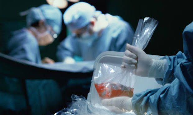
- Step 2: Static Cold Storage (SCS): The most common method. After flushing and removal, organs are placed in sterile bags containing preservation solution and then submerged in ice slush. This maintains the organ at a hypothermic temperature (typically 4-8°C). Different preservation solutions are used depending on the organ (e.g., University of Wisconsin (UW) solution, Histidine-Tryptophan-Ketoglutarate (HTK) solution). Each organ has specific cold ischemic time limits within which transplantation should ideally occur to ensure optimal function.
- Step 3: Machine Perfusion: An increasingly utilized method, particularly for kidneys, but also for livers and lungs. This involves connecting the organ to a machine that perfuses it with a preservation solution (either cold or normothermic, sometimes oxygenated). Machine perfusion can potentially extend preservation times, allow for assessment of organ viability, and potentially improve outcomes compared to SCS, especially for organs from DCD donors or those with marginal function.
- Step 4: Packaging and Transport: Preserved organs are meticulously packaged in sterile containers within insulated transport boxes, clearly labeled with donor identifiers, organ type, and timestamp. Immediate transport to the recipient transplant center is arranged.
Surgical Anatomy of Multi-Organ Donors: The Foundation for Retrieval
Successful multi-organ retrieval requires a detailed understanding of systemic and regional anatomy, particularly the vascular supply and relationships of abdominal and thoracic organs. The goal is to excise organs en bloc (as a connected unit) initially, allowing for detailed separation and preparation on the “back-table” outside the body.
- Key Anatomical Structures for Multi-Organ Retrieval:
- Systemic Vasculature: The aorta (from chest to iliac bifurcation) and inferior vena cava are central. The aorta provides the inflow for flushing; the IVC is vented.
- Celiac Axis: Originating from the aorta, it supplies the liver (hepatic artery), spleen (splenic artery), and stomach (left gastric artery). Careful dissection is needed to preserve the common hepatic artery and its branches to the liver.
- Superior Mesenteric Artery (SMA): Supplies the small intestine and parts of the large intestine. Dissection involves identifying its origin just below the celiac axis.
- Inferior Mesenteric Artery (IMA): Supplies the distal large intestine. Its origin is lower on the aorta.
- Renal Arteries and Veins: Supply the kidneys. Multiple arteries/veins are common and must be identified. The renal veins drain into the inferior vena cava.
- Portal Vein: Formed by the confluence of the superior mesenteric and splenic veins, draining blood from the digestive tract, spleen, and pancreas to the liver. It runs posterior to the pancreas head.
- Pancreas: Lies transversely across the posterior abdominal wall. Retrieval requires careful dissection to preserve its vascular supply (from celiac and SMA branches) and the pancreatic duct. Its close relationship to the portal vein and surrounding retroperitoneal structures is critical.
- Liver: Located in the upper right quadrant. Ligaments holding it in place (falciform, triangular, coronary) must be divided. Its vascular supply (hepatic artery, portal vein, hepatic veins draining into IVC) and biliary anatomy are paramount.
- Kidneys: Located retroperitoneally. Their arterial supply from the aorta and venous drainage into the IVC are key. Careful dissection of ureters down to the bladder is required.
- Thoracic Organs (Heart, Lungs): Require separate dissection but are often removed en bloc in a combined procedure. Anatomy involves the aorta (ascending, arch, descending), pulmonary artery, pulmonary veins, superior vena cava, and bronchi.
The retrieval surgery is a highly coordinated procedure, often involving multiple surgical teams working simultaneously on different organ systems (e.g., one team for abdomen, one for chest). Precise anatomical dissection, often involving removing organs with generous lengths of associated vessels and connective tissue en bloc, ensures the integrity and viability of each organ for transplantation.
Multi-Organ Retrieval Procedure: Deceased Donors (DBD vs. DCD)
While the general steps of multi-organ retrieval share similarities, there are key differences between procedures for DBD and DCD donors, primarily related to the timing of circulatory cessation and subsequent ischemic injury.
- General Steps (Common to DBD and DCD after Declaration of Death and Consent):
- Step 1: Preparation: Donor is brought to the operating room, positioned supine. A wide sterile prep is performed. Surgical instruments, preservation solutions, ice slush, and back-table dissection equipment are prepared.
- Step 2: Incision and Access: A long midline incision is typically made from the sternal notch down to the pubis to allow access to both thoracic and abdominal cavities if needed.
- Step 3: In Situ Assessment: The surgical teams visually inspect the organs in their anatomical position to perform a final assessment of their macroscopic appearance and suitability.
- Step 4: Dissection: Careful dissection is performed to identify and mobilize the key anatomical structures described above (aorta, vena cava, major vessels, ligaments, ureters, bronchi, etc.), preparing for the en bloc removal and cannulation.
- Step 5: Cannulation and Flushing: Cannulas are inserted into the aorta (usually infrarenal) and the inferior vena cava (or superior vena cava). The systemic administration of heparin is performed to prevent clotting. Cold preservation solution is infused rapidly through the aortic cannula, while the venous system is vented via the IVC cannula (or by opening the IVC) to allow blood and perfusate outflow. This step rapidly cools the organs.
- Step 6: Cross-Clamping: The aorta is cross-clamped (usually supra-celiac) to ensure all cold perfusate flows into the abdominal and thoracic organ circulations and to isolate these organs.
- Step 7: Excision and Removal: Organs or organ blocks are systematically excised. The order of removal can vary but often involves collaboration between thoracic and abdominal teams. The organs are placed in sterile bags with cold preservation solution.
- Step 8: Back-Table Dissection: Once removed from the donor, the organs are transferred to a sterile back-table where they are meticulously dissected from the en bloc block, prepared for implantation (trimming excess tissue, preparing vessels, flushing further if needed), and packaged for transport.
- Specifics for DBD Retrieval:
- Circulation and ventilation are maintained until surgically interrupted by the aortic cross-clamp and flushing.
- The surgical team controls the timing of the start of cold ischemia by applying the cross-clamp.
- Warm ischemic time is typically negligible or very short (only the time between cross-clamping and initiation of cold perfusion).
- Specifics for DCD Retrieval:
- The procedure follows the declaration of death based on circulatory criteria after withdrawal of life support.
- Warm Ischemic Time (WIT): The critical difference. WIT begins from the moment circulation ceases until the organs are rapidly cooled with preservation solution. This time must be minimized as much as possible (often targeting <20-30 minutes of functional WIT for many organs).
- Rapid Cooling: The surgical team must be poised to begin rapid in situ flushing immediately after declaration of death.
- Organ Selection: Organs particularly sensitive to WIT, such as the heart, lungs, and often the liver, may not be suitable from DCD donors if the WIT is prolonged. Kidneys are generally more tolerant but outcomes can still be affected.
- Assessment: Organs from DCD donors, especially kidneys and livers, may benefit from or require machine perfusion to assess viability and potentially recover from WIT injury before transplantation.
Assessment and Treatment of Living Donors
Living donation, primarily of a kidney or a segment of the liver, offers an alternative path to transplantation and provides recipients with organs that often have superior long-term outcomes compared to deceased donor organs. The assessment process is rigorous, prioritizing the safety and well-being of the donor above all else.
- Step 1: Initial Screening: Basic medical history, blood type testing, and preliminary health questions to identify obvious contraindications.
- Step 2: Comprehensive Medical Evaluation: This is an extensive evaluation over several days or visits involving:
- Detailed History and Physical Examination: Review of all medical conditions, surgeries, medications.
- Laboratory Tests: Complete blood count, comprehensive metabolic panel (renal and liver function), urinalysis, infectious disease screening (HIV, Hepatitis B/C, etc.), tissue typing (HLA matching), and cross-matching with the potential recipient.
- Imaging Studies: CT angiography of the abdomen/pelvis (for kidney) or abdomen (for liver) to map the vascular and anatomical structure of the organ to be donated. Chest X-ray, potentially other scans to screen for malignancy.
- Cardiovascular Assessment: EKG, often stress testing or echocardiogram, to ensure the donor’s heart can withstand surgery.
- Other Consultations: Depending on history, may include pulmonary function tests, colonoscopy, mammography, etc., as part of general health screening.
- Goal: Ensure the donor is in excellent health, has normal function of the organ to be donated and the remaining organ (e.g., the other kidney), and has no underlying conditions that would be exacerbated by surgery or single-organ status, or that could transmit disease to the recipient.
- Step 3: Psychosocial Evaluation: A crucial step involving interviews with social workers and/or psychologists. This assesses:
- Understanding of the risks and benefits of donation.
- Motivation for donating (ensuring it is voluntary and free of coercion).
- Psychological stability and coping mechanisms.
- Adequacy of support systems for recovery.
- Potential financial or social implications.
- Step 4: Independent Donor Advocate/Ethics Review: Often involves an independent physician or ethics committee reviewing the case to ensure the donor’s interests are protected and the donation is voluntary and medically appropriate.
- Step 5: Final Approval: The multidisciplinary living donor team reviews all assessment data and collectively decides if the potential donor is medically and psychosocially suitable.
- Step 6: Surgical Procedure (Treatment):
- Minimally invasive laparoscopic surgery is the standard for most living kidney donations and increasingly used for liver segments where appropriate. This involves small incisions. Open surgery may still be used in some cases.
- The surgical team carefully dissects and removes the specified organ (one kidney or a liver segment) while preserving the surrounding structures and remaining organ function.
- Pain management, fluid balance, and monitoring of vital signs are critical during and immediately after surgery.
- Step 7: Post-operative Care (Treatment):
- Focuses on pain management, monitoring of recovery of the remaining organ (e.g., contralateral kidney function), monitoring for surgical complications (bleeding, infection, wound healing), and early mobilization.
- Psychological support is continued.
- Donor is discharged once stable and recovering well.
- Step 8: Long-Term Follow-up (Treatment): Essential to monitor the donor’s health for life. This includes regular check-ups, blood tests (e.g., kidney or liver function), blood pressure monitoring, and screening for potential long-term complications or health issues.
Conclusion
The journey from a potential organ donor to a successful transplant recipient is a complex and delicate process requiring the coordinated efforts of numerous medical professionals and support staff. Meticulous donor management optimizes organ quality, while sophisticated preservation techniques minimize ischemic damage. Precise surgical anatomy and technique are fundamental to successful retrieval from deceased donors, with critical differences in approach depending on whether donation follows brain death or circulatory death. For living donors, a comprehensive assessment prioritizes donor safety and well-being, followed by expert surgical and post-operative care, including essential long-term follow-up. Each step in this continuum is vital to honoring the gift of donation and providing life-saving opportunities for transplantation.


