Diagnosing liver diseases requires a systematic and multi-modal approach, integrating clinical presentation with various diagnostic studies. The liver performs numerous vital functions, and its pathology can be subtle or devastating, often presenting with non-specific symptoms until significant damage has occurred. Therefore, selecting the appropriate diagnostic tools is crucial for identifying the underlying cause, assessing the severity of the disease, monitoring progression, and guiding treatment.
Professionals rely on a suite of biochemical, serological, imaging, and sometimes histopathological studies to build a complete picture of a patient’s liver health.
Initial Assessment: Biochemical and Serological Studies
Blood tests are typically the first step in evaluating suspected liver disease. They provide insights into liver function, the presence of injury or inflammation, and can often pinpoint specific etiologies.
- Liver Function Tests (LFTs): This panel commonly includes:
- Alanine Aminotransferase (ALT) and Aspartate Aminotransferase (AST): These enzymes are primarily markers of hepatocyte injury. Elevated levels suggest inflammation or damage to liver cells. ALT is generally more specific to the liver than AST. High levels (>10x upper limit of normal) may suggest acute viral hepatitis, ischemic liver injury, or drug-induced liver injury. Moderately elevated levels can be seen in chronic hepatitis, fatty liver disease, or biliary obstruction. The AST/ALT ratio can sometimes provide clues (e.g., ratio > 2 is often seen in alcoholic liver disease).
- Alkaline Phosphatase (ALP): Primarily associated with the bile ducts, elevated ALP suggests cholestasis (impaired bile flow) or infiltrative liver diseases (e.g., tumors, granulomas). It can also be elevated in bone disorders, so GGT is often measured concurrently.
- Gamma-Glutamyl Transferase (GGT): Also involved in bile metabolism, GGT is sensitive to liver disease and bile duct obstruction. Elevated GGT alongside elevated ALP strongly suggests a hepatobiliary origin for the high ALP. GGT is also inducible by alcohol and certain medications (e.g., phenytoin), making it a less specific but sensitive marker.
- Bilirubin (Total and Direct/Conjugated): Bilirubin is a byproduct of heme metabolism. Elevated total bilirubin can indicate increased production, impaired uptake, conjugation, or excretion. Elevated direct/conjugated bilirubin specifically points towards impaired excretion, often due to hepatocellular dysfunction or cholestasis.
- Tests of Synthetic Function: These tests assess the liver’s ability to synthesize key proteins. They are crucial indicators of chronic liver disease severity and prognosis.
- Albumin: A major protein synthesized by the liver, albumin transports various substances. Low albumin levels can reflect chronic liver disease (as the liver loses synthetic capacity), malnutrition, or protein loss elsewhere (e.g., kidneys, gut). Due to its long half-life (approx. 20 days), it’s not a good marker of acute liver failure.
- Prothrombin Time (PT) / International Normalized Ratio (INR): The liver synthesizes clotting factors. Impaired synthesis leads to a prolonged PT/elevated INR. This is a sensitive indicator of acute liver failure or severe chronic liver disease. Unlike albumin, the shorter half-lives of clotting factors make PT/INR responsive to changes in liver function within hours to days.
- Serological Studies for Specific Etiologies: These tests are vital for identifying the cause of liver disease.
- Viral Hepatitis Panel: Tests for Hepatitis A (HAV IgM/IgG), Hepatitis B (HBsAg, HBsAb, HBcAb, HBeAg, HBeAb, HBV DNA), and Hepatitis C (HCV Ab, HCV RNA). These are essential for diagnosing acute and chronic viral hepatitis, which are major causes of liver disease worldwide.
- Autoimmune Markers: Antinuclear Antibodies (ANA), Anti-Smooth Muscle Antibodies (ASMA), Anti-Liver Kidney Microsome Antibodies (Anti-LKM1), Anti-Mitochondrial Antibodies (AMA). These help diagnose Autoimmune Hepatitis (AIH) and Primary Biliary Cholangitis (PBC).
- Metabolic Markers: Serum Ferritin and Transferrin Saturation (for Hemochromatosis), Serum Ceruloplasmin and Urinary Copper (for Wilson’s Disease), Alpha-1 Antitrypsin levels and Phenotyping (for Alpha-1 Antitrypsin Deficiency). These tests identify genetic/metabolic causes.
Selection Strategy: Initial assessment typically includes a standard LFT panel and sometimes synthetic function tests. Based on clinical suspicion (e.g., risk factors for viral hepatitis, family history), specific serological or metabolic tests are ordered to identify the underlying cause.
Imaging Studies
Imaging techniques provide crucial information about the liver’s size, shape, texture, the presence of masses or lesions, patency of blood vessels, and the presence of complications like ascites or portal hypertension.
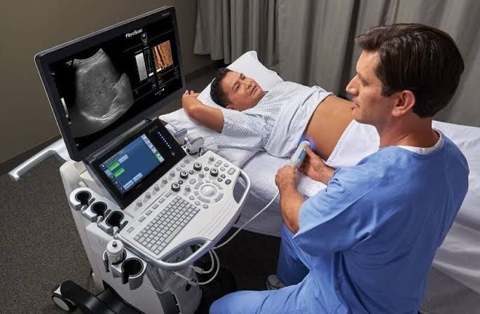
- Ultrasound (US): Often the first imaging modality due to its non-invasiveness, availability, and lower cost. Useful for:
- Assessing liver size and echotexture (e.g., bright echotexture suggests fatty liver, nodular surface suggests cirrhosis).
- Detecting gallstones and bile duct dilatation (indicating obstruction).
- Identifying large masses or cysts.
- Assessing portal and hepatic vein patency using Doppler US.
- Guiding interventional procedures (e.g., biopsy, fluid drainage).
- Limitations: Operator dependent, limited views in obese patients or those with bowel gas, difficulty characterizing small lesions.
- Computed Tomography (CT) Scan: Provides detailed cross-sectional images, often with intravenous contrast. Useful for:
- Characterizing lesions seen on ultrasound.
- Detecting and staging liver tumors (Hepatocellular Carcinoma – HCC, metastases).
- Assessing vascular anatomy and patency more clearly than US.
- Evaluating abdominal organs for extrahepatic causes or complications.
- Limitations: Uses ionizing radiation, contrast carries risks (allergic reaction, kidney function).
- Magnetic Resonance Imaging (MRI): Offers excellent soft tissue contrast and can differentiate between different tissue types using various sequences. Often used for:
- Further characterization of lesions, especially small ones or those difficult to assess on CT.
- Specific sequences (e.g., liver-specific contrast agents) can improve detection of subtle lesions, including HCC.
- MR Cholangiopancreatography (MRCP) specifically visualizes the bile ducts without contrast injection, useful for complex biliary issues.
- Limitations: More expensive and time-consuming than US or CT, availability may be limited, contraindications include certain metallic implants.
- Elastography (e.g., Transient Elastography, ARFI, MRE): Non-invasive techniques that measure liver stiffness, which correlates well with the degree of fibrosis. Increasingly used to:
- Assess the stage of fibrosis in chronic liver diseases (e.g., Hepatitis C, NASH) without biopsy.
- Monitor disease progression or response to treatment.
- Reduce the need for liver biopsy in many patients.
- Limitations: Can be inaccurate in obese patients, those with ascites, or acute inflammation; not useful for diagnosing specific etiologies or characterizing focal lesions.
Selection Strategy: Ultrasound is often the initial imaging study. If US reveals suspicious findings or if the underlying cause diagnosed by labs (e.g., chronic Hepatitis C, NASH) requires fibrosis assessment, elastography may be used. If a mass is suspected or more detailed anatomical information is needed, contrast-enhanced CT or MRI is performed, with MRI often preferred for lesion characterization.
Liver Biopsy: Indications, Contraindications, and Complications
Despite advances in non-invasive tests, liver biopsy remains a valuable tool, particularly when other tests are inconclusive or when detailed histological information is required for diagnosis, prognosis, or treatment planning.
Indications:
- Diagnosis of unexplained abnormal LFTs.
- Staging of fibrosis and grading of inflammatory activity in chronic liver diseases (though non-invasive methods are reducing the need).
- Diagnosis of specific liver diseases when non-invasive tests are insufficient:
- Suspected Autoimmune Hepatitis (AIH).
- Suspected Primary Biliary Cholangitis (PBC) or Primary Sclerosing Cholangitis (PSC) when serology/imaging are atypical.
- Diagnosis and staging of Non-Alcoholic Steatohepatitis (NASH).
- Diagnosis of metabolic disorders (e.g., Hemochromatosis, Wilson’s Disease) when genetic tests are inconclusive or to assess damage.
- Investigation of suspected drug-induced liver injury.
- Evaluation of liver transplant rejection or dysfunction.
- Characterization of hepatic masses or infiltrative diseases (e.g., lymphoma, sarcoidosis, amyloidosis).
- Evaluation of fevers of unknown origin with suspected liver involvement.
Contraindications:
- Absolute:
- Severe uncorrectable coagulopathy (e.g., INR > 1.5-2.0, platelet count < 50,000-60,000/µL). Transjugular approach may be an alternative.
- Severe thrombocytopenia (< 50,000/µL) that cannot be corrected.
- Lack of patient cooperation (requires conscious sedation or general anesthesia alternative).
- Suspected vascular lesion (e.g., hemangioma) due to high bleeding risk.
- Extrahepatic biliary obstruction requiring decompression.
- Relative:
- Moderate ascites (increases risk of bleeding or bile leak; large volume paracentesis before biopsy may help).
- Severe anemia (risk of significant consequences from even minor bleeding).
- Amyloidosis with significant vascular involvement (increased risk of bleeding).
- Skin infection at the biopsy site.
- Morbid obesity (technical difficulty).
- Significant pleural effusion or lung disease (increased risk of pneumothorax).
Complications:
Liver biopsy is generally safe but carries risks. The most common complications include:
- Pain: The most frequent complication, usually localized to the biopsy site or referred to the right shoulder (diaphragmatic irritation), typically manageable with analgesia.
- Bleeding: The most serious potential complication. Can range from minor bruising to significant intrahepatic hematoma or life-threatening hemoperitoneum. Risk is increased with coagulopathy, ascites, or certain underlying liver diseases.
- Pneumothorax/Hemothorax: Risk of puncturing the pleura or lung, usually when the biopsy is performed too high.
- Bile Leak/Peritonitis: Rare, especially with percutaneous biopsy, but possible if a bile duct is punctured. More common with obstructive jaundice.
- Perforation of Other Organs: Very rare (e.g., colon, kidney, gallbladder).
- Infection: Very rare.
- Mortality: Extremely rare (< 0.01%).
Pathological Features of Common Liver Diseases
The histopathological examination of a liver biopsy provides definitive diagnostic information by allowing direct visualization of cellular and architectural changes.
- Viral Hepatitis (Chronic Hepatitis B or C):
- Features: Portal and lobular inflammation (lymphocytes, plasma cells), hepatocyte injury (ballooning, apoptosis/acidophil bodies), steatosis (especially HCV genotype 3), interface hepatitis (inflammation extending from portal tracts into the lobule), and varying degrees of fibrosis, potentially progressing to cirrhosis (bridging fibrosis, nodule formation). Grading (inflammation) and staging (fibrosis) are key components of the report.
- Alcoholic Liver Disease (ALD):
- Features: Steatosis (fat accumulation, often macrovesicular), hepatocyte ballooning degeneration (swollen hepatocytes), Mallory-Denk Bodies (cytoplasmic inclusions of aggregated protein), neutrophilic infiltration (especially in alcoholic hepatitis), and fibrosis, often pericellular (“chicken wire”) pattern, progressing to cirrhosis.
- Non-Alcoholic Fatty Liver Disease (NAFLD) / Non-Alcoholic Steatohepatitis (NASH):
- Features: Spectrum ranging from simple steatosis (fat accumulation) to NASH. NASH shows steatosis, lobular inflammation, hepatocyte ballooning, and often pericellular fibrosis. Histologically similar to AD, but occurs in patients without significant alcohol intake, often associated with metabolic syndrome. Fibrosis staging is critical as it predicts prognosis.
- Autoimmune Hepatitis (AIH):
- Features: Prominent interface hepatitis with a dense infiltrate rich in plasma cells, emperipolesis (lymphocytes within hepatocytes), hepatocyte rosette formation, and varying degrees of fibrosis. Can present acutely or chronically.
- Primary Biliary Cholangitis (PBC):
- Features: Inflammation and destruction of small-to-medium sized bile ducts in the portal tracts (“florid duct lesion”), granuloma formation, varying degrees of interface hepatitis, and progressive fibrosis starting in the portal tracts and leading to cirrhosis. Elevated AMA serology is often present.
- Cirrhosis:
- Features: Defined by architectural distortion characterized by diffuse nodular regeneration surrounded by fibrous septa. Represents the end-stage of many chronic liver diseases. Pathological features also include the underlying cause (e.g., steatosis if due to NASH, iron if due to hemochromatosis).
- Hepatocellular Carcinoma (HCC):
- Features: Malignant proliferation of hepatocytes, typically arising in a background of cirrhosis. Histological patterns vary (trabecular, pseudoglandular, solid). Cytological features include increased nucleus-to-cytoplasm ratio, nuclear pleomorphism, prominent nucleoli, and often increased mitotic activity. Specific stains (e.g., glypican-3, HepPar1) can aid diagnosis.
Conclusion
The diagnosis of liver diseases is a complex but systematic process. It begins with biochemical and serological tests to evaluate liver function, assess injury, and screen for common causes. Imaging studies provide structural information, assess complications, and help characterize lesions. Liver biopsy, while becoming less frequent for staging due to non-invasive methods like elastography, remains invaluable for diagnosing specific conditions, assessing disease activity, and when other methods are inconclusive. Finally, understanding the key pathological features allows for definitive diagnosis and guides patient management. Integrating the findings from all these modalities within the clinical context is essential for accurate diagnosis and effective care of patients with liver disease.


