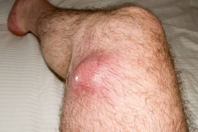
Soft tissue tumors represent a diverse group of neoplasms arising from non-epithelial extraskeletal tissues excluding the central nervous system, hematopoietic system, and lymphoid tissues. This category includes tumors originating from fat, muscle, fibrous tissue, blood vessels, lymphatic vessels, and peripheral nerves. While many soft tissue tumors are benign and pose little threat beyond local symptoms, others are malignant (sarcomas) and can be aggressive, requiring complex management. Understanding their nature, classification, and the diagnostic importance of microscopic examination is crucial for accurate diagnosis, prognosis, and treatment planning.
Classification of Soft Tissue Tumors
The classification of soft tissue tumors is complex and constantly evolving, driven by advancements in molecular biology and pathology. The World Health Organization (WHO) Classification of Tumours is the globally recognized standard. This classification primarily groups tumors based on their presumed line of differentiation or origin tissue, followed by categorization based on behavior (benign, intermediate, or malignant).
Here is a breakdown of the main categories based on tissue differentiation, as outlined by the WHO:
- Adipose Tissue Tumors:
- Benign: Lipoma (very common), Lipomatosis, Hibernoma.
- Intermediate (Locally Aggressive/Rarely Metastasizing): Atypical Lipomatous Tumor/Well-Differentiated Liposarcoma (often locally aggressive, rarely metastasizes unless it dedifferentiates).
- Malignant: Liposarcoma (Myxoid, Round Cell, Dedifferentiated, Pleomorphic subtypes).
- Fibroblastic and Myofibroblastic Tumors:
- Benign: Nodular Fasciitis, Myositis Ossificans, Fibroma of tendon sheath, Elastofibroma.
- Intermediate (Locally Aggressive): Desmoid Tumor (Deep Fibromatosis) – infiltrative locally, high recurrence but typically does not metastasize.
- Intermediate (Rarely Metastasizing): Solitary Fibrous Tumor (can be benign, malignant, or intermediate).
- Malignant: Fibrosarcoma (now less common, diagnosis of exclusion), Myxofibrosarcoma, Low-grade Fibromyxoid Sarcoma, Sclerosing Epithelioid Fibrosarcoma.
- Skeletal Muscle Tumors:
- Benign: Rhabdomyoma (rare in adults, more common in heart of children – often associated with Tuberous Sclerosis).
- Malignant: Rhabdomyosarcoma (primarily a tumor of children and adolescents, but can occur in adults – Embryonal, Alveolar, Pleomorphic, Spindle Cell/Sclerosing subtypes).
- Smooth Muscle Tumors:
- Benign: Leiomyoma (very common, especially in the uterus – fibroids). Can occur in skin, gastrointestinal tract, and deep soft tissues.
- Malignant: Leiomyosarcoma (can arise from smooth muscle in various locations, including blood vessel walls, uterus, retroperitoneum, and skin).
- Vascular Tumors:
- Benign: Hemangioma (capillary, cavernous, venous, arteriovenous, etc. – very common), Lymphangioma, Glomus Tumor.
- Intermediate (Locally Aggressive): Kaposiform Hemangioendothelioma, Retiform Hemangioendothelioma.
- Intermediate (Rarely Metastasizing): Hemangioendothelioma (various subtypes like Epithelioid Hemangioendothelioma).
- Malignant: Angiosarcoma (aggressive, often presenting in skin, soft tissue, breast, or liver).
- Peripheral Nerve Sheath Tumors:
- Benign: Schwannoma (Neurilemoma), Neurofibroma (plexiform neurofibroma is often associated with NF1), Perineurioma.
- Malignant: Malignant Peripheral Nerve Sheath Tumor (MPNST) – often arises from pre-existing neurofibroma, especially in patients with Neurofibromatosis Type 1 (NF1).
- Chondro-osseous Tumors (primarily bone/cartilage, but can present in soft tissue):
- Benign: Extraskeletal Chondroma, Extraskeletal Osteoma.
- Malignant: Extraskeletal Chondrosarcoma, Extraskeletal Osteosarcoma (rare).
- Tumors of Uncertain Differentiation: This is a large and heterogeneous group for tumors that don’t clearly fit established lines of differentiation.
- Benign: Tenosynovial Giant Cell Tumor (Localized and Diffuse types), Giant Cell Tumor of Soft Tissue.
- Intermediate (Locally Aggressive): Aggressive Angiomyxoma.
- Intermediate (Rarely Metastasizing): Synovial Sarcoma (despite the name, it doesn’t arise from synovium, but has a characteristic molecular alteration t(X;18)), Epithelioid Sarcoma.
- Malignant: Alveolar Soft Part Sarcoma, Clear Cell Sarcoma of Soft Parts, Extraskeletal Myxoid Chondrosarcoma (despite name, soft tissue origin), Desmoplastic Small Round Cell Tumor.
- Undifferentiated and Unclassified Sarcomas: These are malignant tumors where the differentiation is not discernible by standard histological and immunohistochemical techniques.
- Malignant: Undifferentiated Pleomorphic Sarcoma (UPS) – formerly known as Malignant Fibrous Histiocytoma (MFH), this is often a diagnosis of exclusion for high-grade pleomorphic sarcomas that lack specific differentiation.
This classification highlights the vast diversity within soft tissue tumors and underscores why accurate diagnosis is paramount. The behavior of these tumors ranges from harmless entities easily removed to highly aggressive malignancies requiring systemic therapy and complex surgical approaches.
Importance of Cytologic and Histologic Features in Diagnosis and Behavior
The definitive diagnosis and assessment of behavior (benign vs. malignant, grade) for soft tissue tumors relies heavily on microscopic examination of tissue or cells obtained through biopsy or surgical excision. Cytology involves examining individual cells, often from fine needle aspiration, while histology involves examining tissue architecture and cell morphology from a core biopsy or surgical specimen.
Pathologists evaluate numerous cytologic and histologic features to arrive at a diagnosis. These features provide crucial clues about the tumor’s origin, specific type, and potential aggressiveness:
- Cellularity: The number of cells per unit area. High cellularity can suggest malignancy.
- Cell Morphology/Cytologic Atypia:
- Nuclear size, shape, and uniformity (Pleomorphism): Variation in nuclear size and shape (pleomorphism) and large, hyperchromatic (dark-staining) nuclei are hallmarks of cytologic atypia, strongly suggestive of malignancy.
- Chromatin pattern: Coarse or vesicular chromatin patterns can be informative.
- Nucleoli: Prominent nucleoli suggest increased metabolic activity, often seen in malignant cells.
- Mitotic Activity: The number of cells undergoing division. A high mitotic rate is a strong indicator of malignancy and is a key criterion for grading sarcomas.
- Necrosis: Areas of cell death within the tumor. Necrosis, especially if not due to biopsy procedures, is often associated with aggressive malignant tumors.
- Growth Pattern: How cells are arranged (e.g., fascicles, sheets, nests, alveolar pattern, myxoid background, storiform pattern). This helps identify the lineage (e.g., fascicles of spindle cells in smooth muscle or fibrous tumors, alveolar pattern in some sarcomas).
- Stromal Features: The appearance of the surrounding connective tissue (stroma). Features like hyalinization, myxoid change (gelatinous background), or inflammatory infiltrate can aid diagnosis.
- Infiltrative vs. Circumscribed Margins: Benign tumors are often well-circumscribed or encapsulated, allowing easy surgical removal. Malignant tumors are frequently infiltrative, invading surrounding normal tissues, making complete removal challenging.
- Ancillary Techniques:
- Immunohistochemistry (IHC): Staining tissue sections with antibodies specific to certain proteins expressed by cells. This is invaluable for confirming lines of differentiation (e.g., desmin for muscle, S100 for nerve sheath or melanoma, CD34 for vascular or some stromal tumors, keratin for epithelial markers seen in some sarcomas like synovial sarcoma).
- Molecular Testing: Detecting specific genetic alterations (e.g., translocations, mutations, gene amplifications) that are characteristic of certain tumor types (e.g., t(X;18) EWSR1-SS18 fusion in Synovial Sarcoma, FUS-DDIT3 fusion in Myxoid Liposarcoma, MDM2 amplification in Atypical Lipomatous Tumor/Well-Differentiated Liposarcoma). Molecular tests are increasingly integrated into the diagnostic process and can be crucial for classification and targeted therapy.
- Cytogenetics/FISH: Analyzing chromosomes or detecting specific gene fusions using fluorescence in situ hybridization.
Collectively, these features allow the pathologist to:
- Identify the specific tumor type: Distinguishing between a lipoma and a liposarcoma, or a benign nerve sheath tumor and an MPNST, is critical.
- Assess the tumor’s behavior: Classifying it as benign, intermediate, or malignant.
- Grade malignant tumors: Sarcoma grading systems (like the FNCLCC system) are based on features such as differentiation, mitotic count, and necrosis, which correlate with the likelihood of metastasis and patient outcome.
- Guide treatment: The specific type and grade dictate whether simple excision is sufficient, or if wider margins, radiation therapy, chemotherapy, or targeted therapy are required.
Therefore, the expertise of a pathologist specializing in soft tissue tumors is paramount. Close collaboration between clinicians (surgeons, oncologists) and pathologists is essential for optimal patient management.
Commonest Benign and Malignant Soft Tissue Tumors
While there are hundreds of types of soft tissue tumors, some are encountered far more frequently than others.
Common Benign Soft Tissue Tumors:
- Lipoma:
- Origin: Adipose tissue.
- Description: The single most common soft tissue tumor. Typically presents as a soft, mobile, painless mass that grows slowly. Commonly found in the subcutaneous tissue of the trunk, neck, and extremities. Histologically, they are composed of mature adipocytes identical to normal fat, usually encapsulated. Removal is typically for cosmetic reasons, symptoms (if pressing on nerves), or diagnostic uncertainty.
- Hemangioma:
- Origin: Vascular tissue.
- Description: Very common, particularly in infants and children (often resolving spontaneously). In adults, they are stable. Can occur anywhere, including skin, subcutaneous tissue, muscle, or internal organs. Histologically, they consist of proliferations of blood vessels. Capillary hemangiomas have small vessels, cavernous hemangiomas have larger, dilated vessels. Often present as reddish or bluish lesions.
- Schwannoma (Neurilemoma) and Neurofibroma:
- Origin: Peripheral nerve sheath.
- Description: Schwannomas are usually solitary and arise from Schwann cells, displacing the nerve fibers. Neurofibromas involve all elements of the nerve (Schwann cells, fibroblasts, axons) and are often multiple, especially in Neurofibromatosis Type 1 (NF1). Usually benign, slow-growing masses, often painful or causing neurological symptoms if compressing the nerve. Histologically, they have specific cellular patterns (Antoni A and B areas often seen in Schwannoma).
- Tenosynovial Giant Cell Tumor (TGCT):
- Origin: Synovial lining of joints, tendon sheaths, and bursae.
- Description: Can be localized (often called giant cell tumor of tendon sheath) presenting as a slow-growing mass on fingers/hands (most common), or diffuse (formerly pigmented villonodular synovitis PVNS) affecting larger joints and causing pain, swelling, and joint destruction. Histologically characterized by multinucleated giant cells, mononuclear cells, and often hemosiderin pigment. Benign or intermediate (locally aggressive).
Common Malignant Soft Tissue Tumors (Sarcomas):
Soft tissue sarcomas are relatively rare cancers, accounting for less than 1% of all adult malignancies. However, within this group, some are more common than others.
- Liposarcoma:
- Origin: Adipose tissue.
- Description: One of the most common sarcomas in adults. Occurs most often in the deep soft tissues of the extremities or retroperitoneum. Has several subtypes with vastly different behaviors: Well-differentiated liposarcoma (often indolent, locally recurrent but rarely metastasizes unless it dedifferentiates), Myxoid/Round cell liposarcoma (distinctive myxoid matrix, often with a specific translocation), Pleomorphic liposarcoma (high-grade, aggressive).
- Leiomyosarcoma:
- Origin: Smooth muscle.
- Description: Can arise from smooth muscle in various locations: uterus (common site), retroperitoneum, skin, and particularly from the walls of blood vessels. Often presents as a firm mass. Histologically shows spindle cells with features of smooth muscle differentiation (e.g., cigar-shaped nuclei). Behavior varies with site and grade; deep-seated and retroperitoneal types are generally high-grade and aggressive.
- Undifferentiated Pleomorphic Sarcoma (UPS):
- Origin: Uncertain, diagnosis of exclusion.
- Description: This is a high-grade sarcoma typically found in the extremities of older adults. It is characterized by highly pleomorphic cells (varied size and shape), numerous mitoses, and often necrosis and hemorrhage. The diagnosis of UPS is made when a high-grade pleomorphic sarcoma lacks sufficient features to classify it into another specific lineage (e.g., poorly differentiated liposarcoma, leiomyosarcoma, rhabdomyosarcoma). It behaves aggressively with a high risk of local recurrence and metastasis.
- Synovial Sarcoma:
- Origin: Uncertain, not related to synovium.
- Description: Despite the name, this sarcoma does not arise from synovial tissue. It is a distinct entity characterized by a specific chromosomal translocation t(X;18) leading to the fusion gene SS18-SSX. Commonly occurs near large joints (but not necessarily within the joint capsule) of the extremities, particularly the knee and ankle, in adolescents and young adults. Can have monophasic (spindle cells) or biphasic (spindle cells and epithelial cells) histological patterns. Tends to be aggressive with a propensity for metastasis.
- Malignant Peripheral Nerve Sheath Tumor (MPNST):
- Origin: Peripheral nerve sheath cells.
- Description: Relatively uncommon but aggressive sarcoma that arises from a peripheral nerve or from a pre-existing benign nerve sheath tumor (neurofibroma or schwannoma). A significant proportion (up to 50%) occur in individuals with Neurofibromatosis Type 1 (NF1). Often presents as a rapidly growing, painful mass along the course of a nerve. Histologically, it’s a spindle cell sarcoma with features of nerve sheath differentiation (sometimes difficult to prove).
Conclusion
Soft tissue tumors represent a vastly heterogeneous group of neoplasms ranging from exceedingly common benign entities like lipoma and hemangioma to rare and aggressive malignancies like liposarcoma and UPS. Their accurate classification is complex and relies fundamentally on detailed microscopic examination – both cytology and histology – often supplemented by immunohistochemistry and molecular testing. The specific features identified by the pathologist directly inform the tumor’s type, its likely biological behavior (benign, intermediate, or malignant), its grade if malignant, and consequently, the necessary diagnostic and therapeutic strategies. Given the complexity and rarity of many soft tissue sarcomas, their management often requires a multidisciplinary team approach involving surgeons, oncologists, radiation therapists, and expert pathologists to ensure optimal patient outcomes. Continued research is essential to further refine classification, improve diagnostic accuracy, and develop more effective treatments for these challenging tumors.


