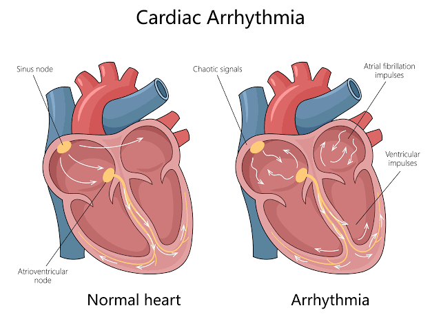
The heart’s electrical system is a complex network responsible for coordinating the rhythmic contraction of the heart chambers, ensuring efficient blood circulation. Disruptions to this system can lead to abnormal heart rhythms (arrhythmias) or impaired impulse transmission (conduction blocks). Understanding the underlying mechanisms and their electrocardiogram (ECG) manifestations is crucial for diagnosis and management.
Mechanisms of Arrhythmias
Arrhythmias often arise from two primary mechanisms: abnormal impulse formation (ectopic foci) or abnormal impulse conduction (re-entry).
- Ectopic Foci of Excitation:
- Definition: An ectopic focus is a site within the heart that initiates an electrical impulse outside of the normal sinoatrial (SA) node, which is the heart’s natural pacemaker.
- Locations: Ectopic foci can be located anywhere in the heart tissue capable of spontaneous depolarization, including:
- Atria (e.g., pulmonary veins, atrial muscle)
- Atrioventricular (AV) junction (junctional tissue)
- Ventricles (e.g., Purkinje fibers, ventricular muscle)
- Mechanism: Ectopic foci can become the dominant pacemaker or interrupt the normal SA node rhythm through several mechanisms:
- Enhanced Automaticity: Cells outside the SA node that normally have latent automaticity (a slower rate of spontaneous depolarization) increase their firing rate, outpacing the SA node. This can occur due to factors like electrolyte imbalances (hypokalemia), hypoxia, ischemia, drug toxicity (e.g., digitalis), or sympathetic stimulation.
- Triggered Activity: Abnormal electrical activity initiated by afterdepolarizations, which are secondary depolarizations that occur during or after the repolarization phase of a cardiac action potential.
- Early Afterdepolarizations (EADs): Occur during phase 2 or 3 of the action potential and can lead to Torsades de Pointes. Often associated with prolonged repolarization (long QT interval).
- Delayed Afterdepolarizations (DADs): Occur after repolarization is complete (phase 4) but before the next normal impulse. Often associated with conditions leading to intracellular calcium overload, such as digitalis toxicity or sympathetic hyperactivity.
- Re-entry Phenomena:
- Definition: Re-entry occurs when an electrical impulse travels through a pathway, and instead of terminating after exciting the downstream tissue, it finds a way to re-excite tissue it has already passed through, setting up a self-perpetuating loop of activation.
- Necessary Conditions for Re-entry: Three conditions are generally required for a re-entrant circuit to form and be maintained:
- Presence of a Circuit: A circular or potential circular pathway for impulse propagation. This can be anatomical (e.g., around a scar, through an accessory pathway) or functional (e.g., based on electrophysiological properties of the tissue).
- Unidirectional Block: Conduction block in one direction within a portion of the circuit, while conduction is possible in the opposite direction. Usually, this means the impulse is blocked anterogradely down one limb of the pathway but can conduct retrogradely up the other limb.
- Slow Conduction: Conduction must be slow enough within the circuit to allow the tissue ahead of the impulse front (that was previously excited) to recover its excitability (repolarize) by the time the impulse returns. This allows the impulse to continuously propagate around the loop.
- Mechanism: An impulse arrives at a point where the pathway splits into two limbs (e.g., alpha and beta pathways in the AV node, or a normal pathway and an accessory pathway). If one limb (say, the beta pathway) is refractory due to a unidirectional block (often related to differing refractory periods), the impulse is forced down the other limb (the alpha pathway). If conduction down the alpha pathway is slow enough, by the time the impulse reaches the end of the alpha pathway and can potentially travel back up the beta pathway, the beta pathway has recovered its excitability from the initial impulse. The impulse then travels retrogradely up the beta pathway, re-enters the origin point, and finds the alpha pathway recovered, allowing the cycle to repeat continuously.
- Common Sites: Re-entry is a common mechanism for many tachyarrhythmias, including:
- AV Nodal Re-entrant Tachycardia (AVNRT) – within the AV node
- AV Re-entrant Tachycardia (AVRT) – involving an accessory pathway (like in Wolff-Parkinson-White syndrome)
- Atrial Flutter – often around the tricuspid annulus
- Ventricular Tachycardia – often around scar tissue from prior infarction
Common Types of Arrhythmias and ECG Appearance
Arrhythmias are broadly classified based on their origin (supraventricular or ventricular) and rate (tachycardia > 100 bpm, bradycardia < 60 bpm). Here we describe common types of tachyarrhythmias:
- Atrial Fibrillation (AFib):
- Description: A supraventricular tachyarrhythmia characterized by chaotic, irregular electrical activity in the atria, leading to ineffective atrial contraction. AV nodal conduction is variable, resulting in an irregularly irregular ventricular rhythm. The primary mechanism is often triggered activity or re-entry originating from foci, particularly around the pulmonary veins, leading to multiple small re-entrant wavelets in the atria.
- ECG Appearance:
- Rate: Atrial rate is very rapid (300-600 bpm) but disorganized. Ventricular rate is typically rapid (>100 bpm) if untreated, but highly variable and irregular.
- Rhythm: Irregularly Irregular (the hallmark feature). No discernible pattern to the R-R intervals.
- P waves: Absent. Replaced by fine to coarse “fibrillatory waves” (f waves) that represent the chaotic atrial activity. These are best seen in leads V1, II, III, and aVF.
- QRS Complex: Usually narrow (<0.12 seconds), as conduction through the ventricles follows the normal His-Purkinje system. QRS shape is generally consistent unless there is a pre-existing bundle branch block or rate-related aberrant conduction.
- P-QRS Relationship: No clear relationship between the fibrillatory activity and the QRS complexes.
- Atrial Flutter (AFlutter):
- Description: A supraventricular tachyarrhythmia caused by a single, consistent re-entrant circuit within the atria, most commonly in the right atrium around the tricuspid annulus (typical counter-clockwise flutter). This produces rapid, regular atrial activation. The AV node then conducts these impulses to the ventricles with a fixed ratio (e.g., 2:1, 3:1, 4:1 block) or variable block.
- ECG Appearance:
- Rate: Atrial rate is rapid and regular, typically 250-350 bpm (classically around 300 bpm for typical flutter). Ventricular rate depends on the degree of AV block (e.g., 150 bpm for 2:1 block, 100 bpm for 3:1 block).
- Rhythm: Atrial rhythm is regular. Ventricular rhythm is regular if there is a fixed AV conduction ratio (e.g., 2:1, 3:1) or irregular if the AV block is variable.
- P waves: Absent. Replaced by characteristic “flutter waves” (F waves) in a “sawtooth” pattern, best seen in leads II, III, and aVF. These represent the rapid, regular atrial depolarization.
- QRS Complex: Usually narrow (<0.12 seconds), similar to AFib, as ventricular conduction is normal.
- P-QRS Relationship: A fixed or variable ratio of F waves to QRS complexes (e.g., 2 F waves for every 1 QRS, 3 F waves for every 1 QRS).
- Supraventricular Tachycardia (SVT):
- Description: An umbrella term for any rapid arrhythmia (>100 bpm) originating above the ventricles. This includes various types, most commonly AV Nodal Re-entrant Tachycardia (AVNRT) and AV Re-entrant Tachycardia (AVRT), but can also include atrial tachycardias. These are often initiated by a premature beat and maintained by a re-entrant circuit.
- ECG Appearance:
- Rate: Rapid, typically 150-250 bpm.
- Rhythm: Usually regular.
- P waves: May be absent, hidden within the QRS complex or the T wave, or occur immediately after the QRS (retrograde P waves). Their appearance and location depend on the specific type of SVT. Often, they are difficult to discern.
- QRS Complex: Usually narrow (<0.12 seconds), as ventricular activation occurs via the normal His-Purkinje system. Wide QRS SVT can occur if there is a pre-existing bundle branch block or rate-related aberrant conduction.
- P-QRS Relationship: Often difficult to determine due to obscured P waves. If visible, retrograde P waves may be seen.
- Ventricular Tachycardia (VT):
- Description: A rapid arrhythmia originating from an ectopic focus or re-entrant circuit within the ventricles. VT is potentially life-threatening as it can compromise cardiac output and degenerate into ventricular fibrillation. It is often associated with structural heart disease, particularly ischemic heart disease leading to scar formation that facilitates re-entry.
- ECG Appearance:
- Rate: Rapid, typically 100-250 bpm.
- Rhythm: Usually regular or slightly irregular.
- P waves: Present or absent. If present, they are typically dissociated from the QRS complexes (atrial and ventricular rhythms are independent). Atrial rate is usually slower than ventricular rate.
- QRS Complex: Wide (>0.12 seconds) and often bizarre in shape, reflecting abnormal, slow ventricular activation outside the normal conduction system. The morphology can be monomorphic (consistent QRS shape, suggesting a single source/circuit) or polymorphic (varying QRS shape, suggesting multiple sources or a constantly changing circuit, such as Torsades de Pointes).
- P-QRS Relationship: Complete AV dissociation is common; ventricular beats are not preceded by P waves. Occasionally, “capture beats” (normal-looking, narrow QRS occurring when a supraventricular impulse manages to conduct through the AV node) or “fusion beats” (a hybrid QRS when a supraventricular and ventricular impulse partially activate the ventricles) may be seen, which help confirm VT.
- Ventricular Fibrillation (VFib):
- Description: A chaotic, life-threatening arrhythmia characterized by disorganized electrical activity in the ventricles, resulting in no coordinated ventricular contraction and thus no effective cardiac output. It is a state of cardiac arrest. The mechanism involves multiple small, erratically moving re-entrant wavelets within the ventricular myocardium.
- ECG Appearance:
- Rate: Indiscernible, chaotic electrical activity.
- Rhythm: Entirely irregular and chaotic.
- P waves: Absent.
- QRS Complex: Absent. Replaced by irregular, undulating waveforms of varying amplitude and shape.
- P-QRS Relationship: No identifiable waveforms or relationships. The tracing appears as a chaotic, wavy line. This rhythm requires immediate defibrillation.
Types of Conduction Block
Conduction blocks occur when the electrical impulse is delayed or completely interrupted as it travels through the heart’s conduction system, most commonly affecting the AV node, Bundle of His, or bundle branches. We will focus on AV blocks here.
- Incomplete Heart Block (AV Blocks): Occur when some, but not all, atrial impulses are conducted to the ventricles, or are conducted with delay.
- First-Degree AV Block:
- Description: A delay in conduction through the AV node. Every atrial impulse is conducted to the ventricles, but it takes longer than normal.
- Mechanism: Slowed conduction velocity primarily within the AV node.
- ECG Appearance:
- Rate & Rhythm: Dependent on the underlying sinus rate and rhythm.
- P waves: Present and regular, each followed by a QRS complex.
- PR Interval: Prolonged and constant, measuring >0.20 seconds in adults.
- QRS Complex: Usually normal width, unless pre-existing bundle branch block is present.
- P-QRS Relationship: A 1:1 relationship between P waves and QRS complexes, with a persistently prolonged PR interval. Generally considered benign, but can be a precursor to higher-degree blocks in some cases.
- Second-Degree AV Block: Occurs when some atrial impulses are blocked and fail to reach the ventricles, resulting in dropped QRS complexes. Two main types exist:
- Mobitz Type I (Wenckebach):
- Description: Characterized by progressive lengthening of the PR interval over several beats until an atrial impulse is completely blocked and a QRS complex is dropped. The cycle then repeats.
- Mechanism: Progressive increase in the refractory period and conduction delay primarily within the AV node, leading to eventual failure of conduction.
- ECG Appearance:
- Rate: Atrial rate (based on P waves) is regular and faster than the ventricular rate (based on QRS complexes). Ventricular rate is irregular.
- Rhythm: Atrial rhythm is regular. Ventricular rhythm is irregular due to the dropped QRS complexes.
- P waves: Present and regular. Not all P waves are followed by a QRS.
- PR Interval: Progressively lengthens with each beat until a P wave is not followed by a QRS. The increment in PR lengthening is typically greatest with the second beat of the cycle. The PR interval after the dropped beat is the shortest in the cycle.
- QRS Complex: Usually normal width, as the block is typically in the AV node. A beat is dropped periodically.
- P-QRS Relationship: A repeating pattern where the PR interval gets longer and longer until a P wave is blocked. The ratio of P waves to QRS complexes is typically n:n-1 (e.g., 3:2, 4:3). Generally considered less serious than Type II.
- Mobitz Type II:
- Description: Characterized by intermittent, sudden drops of QRS complexes without prior progressive lengthening of the PR interval.
- Mechanism: The block is typically below the AV node, usually in the Bundle of His or bundle branches. Conduction is either normal (“all or nothing”) or completely blocked; there’s no progressive delay.
- ECG Appearance:
- Rate: Atrial rate is regular and faster than the ventricular rate. Ventricular rate is usually regular, but slower than the atrial rate, depending on the conduction ratio (e.g., 2:1, 3:1).
- Rhythm: Atrial rhythm is regular. Ventricular rhythm is regular (if the block ratio is fixed, e.g., 2:1) or irregular (if the block ratio varies).
- P waves: Present and regular. More P waves than QRS complexes.
- PR Interval: For the conducted beats, the PR interval is constant and may be normal or prolonged. Crucially, there is no progressive lengthening before a dropped beat.
- QRS Complex: Can be normal or wide depending on whether the block is in the Bundle of His or the bundle branches. A QRS complex is periodically absent after a P wave.
- P-QRS Relationship: A fixed or variable ratio of P waves to QRS complexes (e.g., 2:1 block means every second P wave is blocked). This type is more serious than Mobitz Type I as it is more likely to progress to complete heart block and is often associated with underlying structural disease.
- Mobitz Type I (Wenckebach):
- First-Degree AV Block:
- Complete Heart Block (Third-Degree AV Block):
- Description: A state where no atrial impulses are conducted through the AV node to the ventricles. There is a complete dissociation between atrial and ventricular activity. The ventricles are paced by an escape rhythm originating from below the block site (e.g., AV junction, Bundle of His, or ventricles themselves).
- Mechanism: Complete interruption of conduction at or below the AV node, preventing any atrial impulse from reaching the ventricles.
- ECG Appearance:
- Rate: Atrial rate (P waves) is usually normal or slightly increased. Ventricular rate (QRS complexes) is slow, typically 40-60 bpm if the escape rhythm originates from the junction/His bundle (narrow QRS) or 20-40 bpm if it originates from the ventricles (wide QRS).
- Rhythm: Both atrial and ventricular rhythms are regular, but completely independent of each other. There is no relationship between the timing of P waves and QRS complexes.
- P waves: Present and regular, occurring at their own rate. They march through the QRS complexes independently.
- PR Interval: Variable, constantly changing, as there is no conduction correlation.
- QRS Complex: Width depends on the site of the escape rhythm: narrow if junctional or His bundle, wide if ventricular.
- P-QRS Relationship: Complete AV dissociation. P waves appear at a faster rate than QRS complexes, with no association between them. This is a medical emergency often requiring immediate pacing.
Understanding these mechanisms, characteristic ECG patterns, and classifications of arrhythmias and conduction blocks provides the foundation for interpreting electrocardiograms and managing patients with cardiac electrical disorders. Careful analysis of rate, rhythm, P waves, PR interval, QRS complex, and the relationship between P waves and QRS complexes is essential for accurate diagnosis.


