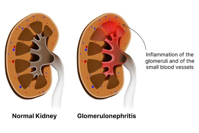
Glomerulonephritis (GN) represents a group of kidney diseases characterized by inflammation and damage to the glomeruli, the tiny filtering units within the kidney. Healthy glomeruli are vital for removing waste products and excess fluid from the blood while retaining essential proteins and cells. When glomeruli are injured, this filtration process is compromised, leading to a range of clinical manifestations and potentially progressive kidney failure. Understanding the mechanisms by which glomerular injury occurs (pathogenesis), how the glomerulus responds to insult, and the resulting clinical patterns is crucial for diagnosis, treatment, and prognosis.
The Pathogenesis of Glomerulonephritis
The pathogenesis of glomerulonephritis is complex and often involves immunological mechanisms, although non-immunological factors can also contribute or exacerbate injury. The primary focus is typically on how the body’s immune system targets the glomerulus.
- Immunological Mechanisms: These are the most common causes of primary glomerulonephritis and involve the deposition or formation of immune complexes within the glomerulus, or direct autoantibody attack.
- Immune Complex-Mediated Injury: This is the most frequent mechanism. Immune complexes (antigen-antibody complexes) can cause damage in two main ways:
- In Situ Formation: Antibodies react directly with antigens that are already present within the glomerulus.
- Anti-Glomerular Basement Membrane (Anti-GBM) Disease: Antibodies target specific components of the glomerular basement membrane (GBM), primarily the alpha3 chain of type IV collagen. Binding of these antibodies activates the complement system (specifically the classical pathway) and attracts inflammatory cells like neutrophils and macrophages. This leads to rapid, severe damage characterized by GBM destruction and often crescent formation. Goodpasture syndrome is a classic example.
- Heymann Nephritis (Experimental Model): Antibodies bind to antigens on the podocyte surface, leading to immune complex formation in situ and subepithelial deposits, characteristic of membranous nephropathy. In human membranous nephropathy, antibodies often target podocyte antigens like the M-type phospholipase A2 receptor (PLA2R).
- Deposition of Circulating Immune Complexes: Pre-formed antigen-antibody complexes circulating in the blood can become trapped within the glomerular filtration barrier based on their size, charge, and affinity for glomerular components.
- Subendothelial Deposits: Often seen in diseases like Post-infectious GN (e.g., post-streptococcal), Lupus Nephritis (Class III or IV), and IgA Nephropathy. These complexes active complement (classical and alternative pathways) and attract inflammatory cells to the capillary lumen and subendothelial space, causing endothelial damage and proliferation.
- Subepithelial Deposits: Typically seen in Post-infectious GN and Membranous Nephropathy. These deposits tend to be larger and activate complement, leading to membrane attack complex formation and podocyte injury.
- Mesangial Deposits: Common in IgA Nephropathy and Lupus Nephritis (Class II). Complexes deposit in the mesangium, stimulating mesangial cell proliferation and matrix expansion.
- Regardless of location, deposited immune complexes activate the complement cascade (classical or alternative pathways), leading to the generation of anaphylatoxins (C3a, C5a) and the membrane attack complex (MAC – C5b-9). Anaphylatoxins are potent chemoattractants for inflammatory cells, while MAC can directly injure glomerular cells. This inflammatory cascade releases cytokines, chemokines, growth factors, proteases, and reactive oxygen species, all contributing to glomerular damage.
- In Situ Formation: Antibodies react directly with antigens that are already present within the glomerulus.
- Cell-Mediated Immunity: While less prominent than immune complex disease in many GN types, T-cells and macrophages play a critical role in amplifying injury and contributing to fibrosis and crescent formation, particularly in severe inflammatory forms like T-cell mediated interstitial nephritis or contributing to the damage in crescentic GN. Sensitized T lymphocytes can directly injure glomerular cells or release cytokines that recruit and activate macrophages.
- Alternative Pathway Complement Dysfunction: In some forms of GN, particularly C3 Glomerulopathy (including Dense Deposit Disease and C3 Glomerulonephritis), the primary defect lies in the dysregulation of the alternative pathway of the complement system. This leads to uncontrolled deposition of C3 breakdown products within the glomerulus without significant immunoglobulin deposition. This excessive complement activation directly damages glomerular structures.
- Immune Complex-Mediated Injury: This is the most frequent mechanism. Immune complexes (antigen-antibody complexes) can cause damage in two main ways:
- Non-Immunological Mechanisms: While less central to the initiation of many forms of GN, non-immunological factors can contribute significantly to the progression of kidney damage, particularly leading to glomerulosclerosis.
- Hyperfiltration and Hypertrophy: In conditions with reduced nephron mass (e.g., due to prior inflammatory injury or other causes), the remaining glomeruli experience increased blood flow and pressure (hyperfiltration). This leads to hypertrophy of glomerular cells, increased mesangial matrix production, podocyte stress, and ultimately, glomerulosclerosis (scarring). This mechanism is central to the progression of many chronic kidney diseases, including those initially triggered by GN.
- Podocyte Injury: Podocytes are critical for maintaining the filtration barrier. Various insults, including immunological attacks, genetic defects, toxins, and mechanical stress (hyperfiltration), can injure podocytes. Podocyte effacement, detachment, and loss are key features leading to heavy proteinuria and contributing to glomerulosclerosis.
- Coagulation Pathways: Activation of coagulation within glomerular capillaries can lead to fibrin deposition, microthrombi, and further injury, particularly seen in conditions like hemolytic uremic syndrome (HUS) or severe vasculitis affecting the glomerulus.
In summary, the pathogenesis of GN is primarily driven by immune responses that deposit or target structures within the glomerulus, triggering inflammatory and cytotoxic cascades. These initial insults are often compounded by non-immunological factors that promote progressive scarring.
Basic Reactions of the Glomerulus to Injury
Regardless of the specific cause, the glomerulus responds to injury through a limited number of stereotypic reactions that represent cellular and matrix alterations aiming to repair or contain the damage. These changes are key pathological features observed in kidney biopsies.
- Cellular Proliferation:
- Endothelial Cell Proliferation: Swelling and increase in number of endothelial cells lining the glomerular capillaries. Often seen in acute inflammatory conditions (e.g., post-infectious GN, lupus nephritis). Can narrow or occlude capillary lumens.
- Mesangial Cell Proliferation: Increase in the number of mesangial cells located in the central part of the glomerulus. A common reaction to various insults (e.g., IgA nephropathy, mesangioproliferative GN). Increased mesangial cells also produce excess mesangial matrix.
- Parietal Epithelial Cell Proliferation (Crescent Formation): Proliferation of the cells lining Bowman’s capsule. This occurs in response to severe capillary damage where plasma proteins and inflammatory cells leak into the urinary space. Accumulated cells and fibrin form a “crescent” that compresses the glomerular tuft, leading to rapid loss of function. Characteristic of Rapidly Progressive Glomerulonephritis (RPGN).
- Leukocyte Infiltration: Inflammatory cells, primarily neutrophils, lymphocytes, and macrophages, are recruited into the glomerular capillaries and mesangium in response to chemotactic factors released during immune activation. These cells contribute to tissue damage through the release of proteases, cytokines, and reactive oxygen species.
- Glomerular Basement Membrane (GBM) Thickening: The GBM may become diffusely or focally thickened. This can be due to:
- Deposition of immune complexes or abnormal proteins (e.g., amyloid, diabetic nephropathy).
- Increased synthesis of GBM components.
- Seen characteristically in Membranous Nephropathy and Diabetic Nephropathy.
- Glomerulosclerosis: This refers to the irreversible scarring and hardening of the glomerulus due to excessive accumulation of extracellular matrix proteins (collagen, fibrin, etc.). It occurs as a final common pathway after chronic or severe injury.
- Focal Sclerosis: Affecting only some glomeruli.
- Segmental Sclerosis: Affecting only a portion of a single glomerulus. Seen in Focal Segmental Glomerulosclerosis (FSGS).
- Global Sclerosis: Affecting the entire glomerular tuft. Glomerulosclerosis leads to loss of filtration function.
- Podocyte Injury/Effacement: Damage to the podocytes, specialized epithelial cells covering the outside of the capillaries, is a critical event leading to proteinuria. Podocyte foot processes normally interdigitate, forming slit diaphragms. Injury causes these processes to flatten and fuse (effacement), disrupting the filtration barrier and allowing proteins (especially albumin) to leak into the urine. Severe podocyte loss contributes to sclerosis.
- Hyalinosis: Accumulation of homogeneous, eosinophilic material within glomerular capillaries or segments. This material consists of plasma proteins trapped in areas of endothelial injury and increased permeability, often associated with sclerosis.
These reaction patterns provide important clues to the type and severity of glomerular injury seen on kidney biopsy.
Renal Syndromes Associated with Renal Pathology
Glomerular and other renal pathologies manifest clinically as distinct patterns of signs and symptoms known as renal syndromes. These syndromes reflect the net effect of the underlying tissue damage on kidney function. While not exclusive to glomerular disease (tubulointerstitial or vascular injury can also contribute), several key syndromes are particularly associated with glomerulonephritis.
Here is a list of different renal syndromes associated with renal pathology, particularly relevant to glomerular disease:
- Nephritic Syndrome: This syndrome is typically associated with acute inflammatory damage to the glomeruli.
- Key Features: Hematuria (often macroscopic, “cola-colored” urine due to red blood cell casts), moderate proteinuria (usually <3.5 g/day), hypertension (due to fluid retention and renal ischemia), azotemia (elevated blood urea nitrogen and creatinine indicating reduced GFR), and oliguria (reduced urine output).
- Pathology Correlation: Reflects inflammatory injury to the glomerular capillaries, disrupting the filtration barrier and causing red blood cells to leak through, while also reducing GFR due to inflammation and cellular proliferation, leading to fluid and waste retention.
- Nephrotic Syndrome: This syndrome is characterized by severe damage to the glomerular filtration barrier, particularly podocyte injury.
- Key Features: Heavy proteinuria (typically >3.5 g/day in adults), hypoalbuminemia (low blood albumin due to protein loss), generalized edema (due to low oncotic pressure from hypoalbuminemia), and hyperlipidemia/lipiduria (complex metabolic derangements).
- Pathology Correlation: Reflects severely increased permeability of the glomerular filtration barrier (primarily affecting podocytes), allowing large amounts of protein, especially albumin, to escape into the urine.
- Rapidly Progressive Glomerulonephritis (RPGN): A clinical syndrome defined by rapid loss of kidney function (typically a 50% decline in GFR within weeks to months).
- Key Features: Presents as a severe nephritic syndrome with prominent azotemia.
- Pathology Correlation: Almost always associated with extensive crescent formation in the glomeruli, indicating widespread and severe glomerular damage and inflammation. Can be caused by various underlying diseases (e.g., anti-GBM disease, severe immune complex GN like post-infectious or lupus nephritis, ANCA-associated vasculitis affecting the kidney).
- Asymptomatic Hematuria and/or Proteinuria: Detection of blood and/or protein in the urine on routine testing without other symptoms or significant impairment of kidney function.
- Key Features: Hematuria (usually microscopic), mild to moderate proteinuria.
- Pathology Correlation: Suggests milder or earlier-stage glomerular injury (e.g., IgA nephropathy, Thin Basement Membrane Disease) or other urinary tract issues. Requires investigation to determine the cause and prognosis.
- Acute Kidney Injury (AKI): A sudden and significant decline in kidney function (rise in serum creatinine and/or decrease in urine output). While AKI has multiple causes (prerenal, intrinsic renal, postrenal), severe or rapidly progressive glomerulonephritis, or acute tubulointerstitial nephritis secondary to inflammation, can present as intrinsic AKI.
- Pathology Correlation: Reflects acute damage to structures involved in filtration and tubular function, potentially due to severe inflammation, necrosis, or blockage within the kidney.
- Chronic Kidney Disease (CKD): A progressive and often irreversible decline in kidney function over months or years, defined by structural or functional abnormalities of the kidney persisting for >3 months.
- Key Features: Progressively increasing serum creatinine, decreasing GFR, abnormalities in urine analysis, electrolyte imbalances, anemia, bone disease, etc., in later stages.
- Pathology Correlation: Represents the end result of chronic kidney damage from various causes, including persistent or recurrent glomerulonephritis leading to widespread glomerulosclerosis, tubulointerstitial fibrosis, and vascular damage. Many forms of glomerulonephritis, if not successfully treated, can progress to CKD. Tubulointerstitial damage frequently accompanies and contributes to progressive CKD in glomerular diseases.
These syndromes represent the clinical spectrum of kidney disease and highlight how different types and severities of damage at the tissue level translate into observable signs and symptoms in patients.
Conclusion
Glomerulonephritis is a significant cause of kidney disease, driven primarily by complex immunological mechanisms that target the glomerular structures. The glomerulus responds to these insults through a limited repertoire of reactions, including cellular proliferation, leukocyte infiltration, basement membrane changes, podocyte injury, and ultimately, sclerosis. These underlying pathological processes manifest clinically as distinct renal syndromes such as the nephritic syndrome, nephrotic syndrome, RPGN, or asymptomatic urinary abnormalities. Understanding the intricate links between the pathogenesis of glomerular injury, the tissue’s response patterns, and the resulting clinical syndromes is fundamental for healthcare professionals involved in the diagnosis and management of patients with kidney disease. Early identification of the specific pathological process is critical to guide targeted therapies and potentially halt or slow the progression to chronic kidney failure.


