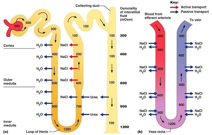
The kidney plays a vital role in maintaining homeostasis by regulating fluid and electrolyte balance. A key function is the ability to produce urine that is either more dilute or more concentrated than plasma. This fine-tuning allows the body to conserve water when dehydrated or excrete excess water when needed. The intricate mechanisms behind this process primarily involve countercurrent systems within the renal medulla, specifically the countercurrent multiplier in the Loop of Henle and the countercurrent exchanger in the vasa recta, significantly aided by the cycling of urea.
The Medullary Osmotic Gradient: The Foundation
- Concept: The ability to concentrate urine relies on the presence of a steep osmotic gradient in the renal medulla. Osmolarity (solute concentration) progressively increases from the corticomedullary border (around 300 mOsm/L, similar to plasma) down to the tip of the renal papilla (reaching up to 1200 mOsm/L or even higher in some species).
- Significance: This hypertonic medullary interstitium acts as a powerful osmotic driving force. As tubular fluid passes through the collecting duct, which traverses this region, water is drawn out of the tubule lumen into the surrounding interstitium by osmosis, thereby concentrating the urine within the collecting duct.
- Components: The two primary solutes contributing to this gradient are sodium chloride (NaCl) and urea. Approximately half of the medullary osmolarity at the papilla is due to accumulated NaCl, and the other half to urea.
The Countercurrent Multiplier: Building the Gradient (Loop of Henle)
- Mechanism: The Loop of Henle functions as a countercurrent multiplier. It uses energy (active transport) in one segment to create an osmotic difference that is then ‘multiplied’ along the length of the loop due to the countercurrent flow and differing permeabilities of the descending and ascending limbs.
- Structure: The loop consists of a descending limb and an ascending limb, with fluid flowing in opposite directions (countercurrent). The ascending limb is further divided into a thin segment and a thick segment.
- Process Steps:
- Fluid Entry: Isosmotic fluid (around 300 mOsm/L) from the proximal tubule enters the descending limb of the Loop of Henle.
- Descending Limb (Permeable to Water, Impermeable to Solutes): As the tubular fluid descends into the increasingly hypertonic medullary interstitium, water moves passively out of the tubule into the interstitium down its osmotic gradient. Solutes, including NaCl and urea, cannot readily enter the tubule here. This outflow of water concentrates the tubular fluid, reaching its highest osmolarity at the bend of the loop (matching the surrounding interstitial osmolarity, e.g., 1200 mOsm/L).
- Thin Ascending Limb (Impermeable to Water, Permeable to Solutes – Passive): As the now concentrated fluid ascends, it enters the thin ascending limb. This segment is impermeable to water. It is, however, passively permeable to NaCl. Driven by the concentration gradient established in the descending limb, NaCl diffuses out of the tubule into the interstitium. This process helps add solutes to the medulla and begins to dilute the tubular fluid.
- Thick Ascending Limb (Impermeable to Water, Permeable to Solutes – Active): The fluid, now slightly less concentrated, moves into the thick ascending limb. This segment remains impermeable to water. Critically, cells in the thick ascending limb actively transport NaCl out of the tubule lumen into the medullary interstitium via the Na+-K+-2Cl- cotransporter (NKCC2) and other transporters. This active transport further adds solutes to the interstitium, maintaining and building the gradient. Because solutes are actively removed while water is retained, the tubular fluid becomes significantly dilute by the time it reaches the distal convoluted tubule (falling to around 100 mOsm/L).
- Multiplication Effect: The key to the “multiplier” is that the single effect (the osmotic difference created by NaCl movement out of the ascending limb) is multiplied along the length of the loop by the continuous flow of fluid. The concentrated fluid arriving at the ascending limb pushes the concentration process further down the loop, amplifying the initial gradient.
The Countercurrent Exchanger: Maintaining the Gradient (Vasa Recta)
- Mechanism: The vasa recta are the peritubular capillaries that dip down into the renal medulla, running parallel to the Loops of Henle and collecting ducts. They function as a countercurrent exchanger, crucial for preserving the medullary osmotic gradient created by the Loop of Henle.
- Structure: These blood vessels also have descending and ascending limbs, with blood flowing in a countercurrent direction relative to the Loop of Henle.
- Process Steps:
- Blood Entry: Blood entering the descending limb of the vasa recta from the cortex has an osmolarity similar to plasma (~300 mOsm/L).
- Descending Vasa Recta: As the blood descends into the increasingly hypertonic medulla, water moves out of the capillaries into the interstitium, and solutes (NaCl and urea) move into the capillaries from the interstitium. This process causes the blood osmolarity to gradually increase, reaching its peak at the bend of the vasa recta (matching the surrounding interstitial osmolarity).
- Ascending Vasa Recta: As the blood ascends back towards the cortex, it passes through regions of decreasing interstitial osmolarity. Now, solutes (NaCl and urea) move out of the capillaries into the interstitium, and water moves into the capillaries from the interstitium.
- Exchanger Effect: Because the blood in the vasa recta equilibrates with the surrounding interstitium at each level of the medulla, the solutes that entered the descending limb are largely returned to the medulla from the ascending limb, and the water that left the descending limb is largely picked up by the ascending limb. This countercurrent exchange minimizes the removal of solutes from the medullary interstitium and prevents the gradient from being “washed away” by blood flow, thus preserving the osmotic driving force for water reabsorption from the collecting duct.
The Crucial Role of Urea
- Contribution to Gradient: Urea is essential for generating the maximal medullary osmotic gradient, particularly in the inner medulla and papilla. While NaCl accounts for significant osmolarity, urea can contribute up to 50% in the deepest regions.
- Urea Cycling: Urea undergoes a complex cycling process within the kidney:
- Filtration: Urea is freely filtered at the glomerulus.
- Proximal Reabsorption: Approximately 50% of filtered urea is passively reabsorbed in the proximal tubule, driven by the movement of water.
- Loop Secretion: Urea is then secreted back into the tubular fluid in the thin descending limb and thin ascending limb of the Loop of Henle.
- Distal/Cortical Collecting Duct Impermeability: The distal convoluted tubule and cortical collecting duct are largely impermeable to urea.
- Inner Medullary Collecting Duct Reabsorption: The permeability of the inner medullary collecting duct to urea is regulated by Antidiuretic Hormone (ADH). ADH increases the insertion of specific urea transporters (UT-A1 and UT-A3) into the apical and basolateral membranes of collecting duct cells in the inner medulla. This allows urea to move out of the tubular fluid down its concentration gradient into the medullary interstitium, significantly contributing to the hyperosmolarity of this region. Urea also enters the interstitium via UT-A2 transporters in the thin ascending limb.
- Importance: The active reabsorption of urea from the inner medullary collecting duct, stimulated by ADH, increases the interstitial osmolarity in the deep medulla. This amplified gradient allows for maximal water reabsorption from the collecting duct under conditions of dehydration, enabling the production of highly concentrated urine. Without efficient urea cycling and regulated reabsorption, the kidney’s capacity to concentrate urine would be significantly reduced.
Dilution of Urine
- Mechanism: Urine dilution occurs when the body has excess water and needs to excrete it. This process relies on removing solutes from the tubular fluid while preventing water reabsorption in later segments.
- Process:
- The active transport of NaCl out of the thick ascending limb renders the tubular fluid dilute (hypotonic, ~100 mOsm/L). This segment is impermeable to water, so water cannot follow the solutes out.
- In the absence of ADH (which occurs when the body is well-hydrated), the collecting duct remains relatively impermeable to water (low expression of aquaporin-2 channels).
- Outcome: As the dilute fluid passes through the collecting duct within the medulla and cortex, very little water is reabsorbed because the tubule is impermeable to water, despite the presence of the medullary gradient. The dilute fluid continues to the renal pelvis and is excreted as hypotonic urine, effectively removing excess water from the body while conserving solutes.
Concentration of Urine
- Mechanism: Urine concentration occurs when the body needs to conserve water (e.g., during dehydration). This process requires the presence of the medullary osmotic gradient and increased water reabsorption.
- Process:
- The medullary osmotic gradient is established and maintained by the countercurrent multiplier (Loop of Henle) and exchanger (vasa recta), with a critical contribution from urea cycling.
- In response to dehydration, the posterior pituitary releases ADH.
- ADH acts on the collecting ducts (and distal tubules), binding to receptors and triggering the insertion of aquaporin-2 water channels into the apical membrane of the principal cells.
- Outcome: The collecting duct becomes highly permeable to water. Driven by the strong osmotic gradient in the medullary interstitium, water moves rapidly out of the tubular fluid, across the tubule cells, and into the interstitium, where it is picked up by the vasa recta. This extensive water removal concentrates the solutes remaining in the tubular fluid. The highly concentrated fluid that remains is excreted as hypertonic urine, allowing the body to conserve water.
In summary, the ability of the kidney to produce both dilute and concentrated urine is a remarkable feat of physiological engineering. It relies on the precise structural arrangement of the Loop of Henle and vasa recta within the renal medulla, enabling countercurrent flow. The countercurrent multiplier actively builds a steep osmotic gradient using the differing permeabilities and transport properties of the Loop of Henle segments. The countercurrent exchanger passively preserves this gradient by minimizing solute removal from the medullary interstitium. The active and regulated reabsorption of urea, particularly from the inner medullary collecting duct under the influence of ADH, is indispensable for achieving the highest levels of urine concentration. Together, these mechanisms allow the kidney to precisely regulate body fluid osmolarity and maintain water balance.


