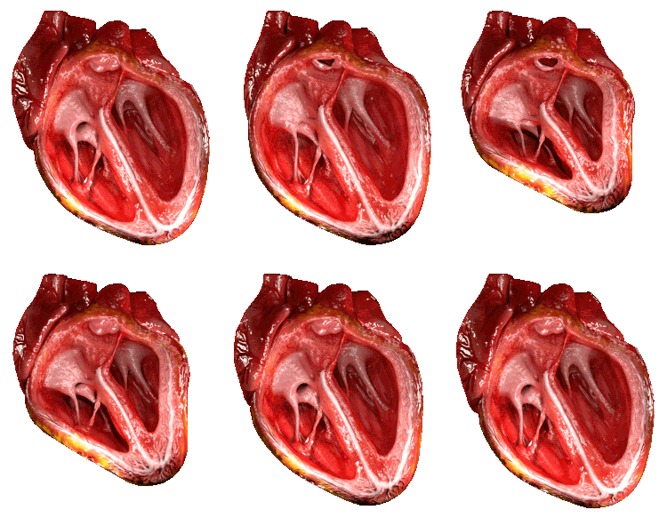
Cardiac pumping, quantified primarily as cardiac output (CO) – the volume of blood pumped by the heart per minute – is a dynamic process essential for circulating oxygenated blood and nutrients throughout the body and removing metabolic waste products. Cardiac output is determined by two key components: stroke volume (SV), the volume of blood ejected by the left ventricle in one contraction, and heart rate (HR), the number of beats per minute (CO = SV x HR). The regulation of these variables is complex and involves a interplay of factors acting both within the heart muscle itself (intrinsic) and from external influences (extrinsic), alongside cellular-level determinants such as ion concentrations and energy availability. Understanding these factors is fundamental to comprehending cardiovascular physiology and pathology.
Intrinsic Regulation of Cardiac Pumping: The Frank-Starling Mechanism
Intrinsic regulation refers to the heart’s inherent ability to adjust its pumping capacity in response to changes in the volume of blood returning to it, independent of external nervous or hormonal control. The primary mechanism for this intrinsic regulation is the Frank-Starling law of the heart, also known as the Starling mechanism.
Explanation of the Frank-Starling Mechanism:
The Frank-Starling law states that, within physiological limits, the stroke volume of the heart increases in response to an increase in the volume of blood filling the heart (end-diastolic volume) when all other factors remain constant. Essentially, the heart pumps out the volume of blood that returns to it.
- Mechanism: As venous return increases, more blood fills the ventricles during diastole, leading to a greater end-diastolic volume. This increased volume stretches the cardiac muscle fibers (myocytes) to a greater extent.
- Cellular Basis: At a myocyte level, this increased stretch brings the actin and myosin filaments within the sarcomeres into a more optimal alignment for cross-bridge formation during subsequent contraction. This increased overlap allows for a greater number of myosin heads to interact with actin binding sites, resulting in a more forceful contraction. Furthermore, stretch increases the sensitivity of the contractile proteins (specifically troponin C) to calcium (Ca++), meaning a given amount of calcium produces a stronger contraction.
- Physiological Importance: The Frank-Starling mechanism is crucial for matching the output of the right ventricle to the output of the left ventricle, preventing blood from backing up in the pulmonary or systemic circulation. For example, if venous return to the right heart increases (e.g., during exercise), the right ventricle’s end-diastolic volume and subsequent stroke volume increase. This increased output from the right ventricle leads to increased filling of the left ventricle, which in turn increases left ventricular end-diastolic volume and stroke volume, thus maintaining balance. It also allows the heart to adapt to varying volumes of venous return without immediate external nervous system intervention.
The Frank-Starling mechanism is a reflection of the inherent contractile properties of the cardiac muscle itself and serves as a fundamental baseline for cardiac output regulation.
Extrinsic Regulation of Cardiac Pumping: The Autonomic Nervous System
Extrinsic regulation involves influences originating from outside the heart, primarily mediated by the autonomic nervous system (ANS) and circulating hormones. The ANS provides rapid and precise control over heart rate and contractility, allowing the cardiovascular system to adapt quickly to changing physiological demands, such as those experienced during exercise, stress, or sleep.
Effect of the Autonomic Nervous System on Heart Pumping:
The ANS consists of two main branches with opposing effects on the heart: the sympathetic nervous system and the parasympathetic nervous system.
- Sympathetic Nervous System:
- Activation: The sympathetic nervous system is often associated with the “fight or flight” response. It is activated during periods of physical activity, stress, or excitement.
- Neurotransmitters: Sympathetic nerves release norepinephrine at the nerve terminals in the heart, which acts on beta-1 adrenergic receptors on cardiac cells (specifically in the sinoatrial (SA) node, atrioventricular (AV) node, atria, and ventricles). The adrenal medulla also releases epinephrine into the bloodstream, which acts on the same receptors with similar effects.
- Effects:
- Increased Heart Rate (Positive Chronotropy): Norepinephrine and epinephrine increase the rate of depolarization of the SA node, leading to a faster heart rate.
- Increased Contractility (Positive Inotropy): They increase the force of contraction of the atrial and ventricular muscle fibers, resulting in a greater stroke volume at any given end-diastolic volume (i.e., shifting the Frank-Starling curve upwards and to the left). This is achieved by increasing calcium influx into the cell and enhancing calcium release from the sarcoplasmic reticulum, making more calcium available for binding to troponin C.
- Increased Conduction Velocity (Positive Dromotropy): They increase the speed of electrical impulse conduction through the AV node and the His-Purkinje system, ensuring rapid and coordinated ventricular contraction.
- Increased Relaxation Rate (Positive Lusitropy): They also accelerate the rate of relaxation of cardiac muscle, allowing for faster filling during diastole, which becomes important at high heart rates.
- Overall Effect: Sympathetic stimulation significantly increases cardiac output by simultaneously increasing both heart rate and stroke volume.
- Parasympathetic Nervous System:
- Activation: The parasympathetic nervous system is associated with the “rest and digest” state. Its influence dominates at rest.
- Neurotransmitter: Parasympathetic nerves (primarily via the vagus nerve) release acetylcholine, which acts on muscarinic (M2) receptors primarily located on cells in the SA node, AV node, and atria. There is less significant parasympathetic innervation to the ventricles, so its effect on ventricular contractility is less pronounced compared to the sympathetic system.
- Effects:
- Decreased Heart Rate (Negative Chronotropy): Acetylcholine decreases the rate of depolarization of the SA node, slowing the heart rate.
- Decreased Conduction Velocity (Negative Dromotropy): It slows the conduction of electrical impulses through the AV node.
- Decreased Atrial Contractility (Negative Inotropy): It slightly decreases the force of contraction in the atria. While it has some direct effect on ventricular function, its primary impact on ventricular output is indirect, resulting from the slower heart rate allowing for more filling time (increasing end-diastolic volume) but also reducing the total number of contractions per minute.
- Overall Effect: Parasympathetic stimulation decreases cardiac output primarily by reducing heart rate.
The ANS exerts tonic (ongoing) influence on the heart, with the parasympathetic system generally dominant at rest, maintaining a heart rate lower than the intrinsic rate of the SA node. Changes in physiological state result in shifts in the balance between sympathetic and parasympathetic activity, allowing for fine-tuning of heart rate and contractility to match the body’s metabolic demands.
Effect of Ions (K+ and Ca++) on Heart Function
Cardiac function, encompassing both electrical activity (generation and conduction of impulses) and mechanical activity (contraction and relaxation), is highly dependent on the precise concentrations and movements of specific ions across the cell membrane. Potassium (K+) and Calcium (Ca++) are particularly critical.
- Calcium (Ca++):
- Role in Contraction: Calcium is the central regulator of cardiac muscle contraction (excitation-contraction coupling). When a cardiac myocyte is depolarized during an action potential, voltage-gated calcium channels open, allowing a small influx of extracellular Ca++. This influx triggers the release of a much larger amount of Ca++ from the sarcoplasmic reticulum (a specialized intracellular calcium store). This surge of intracellular Ca++ binds to troponin C, causing a conformational change in the troponin-tropomyosin complex, which uncovers the myosin binding sites on the actin filaments. Myosin heads can then bind to actin, initiating the cross-bridge cycling that drives muscle contraction.
- Role in Electrical Activity: Calcium influx during phase 2 (plateau phase) of the cardiac action potential is crucial for prolonging the action potential duration and preventing tetanus (sustained contraction). It also plays a role in the slow depolarization of the SA and AV nodes.
- Effect of Altered Levels:
- Hypercalcemia (High Ca++): Increased extracellular calcium increases calcium influx and loading of the sarcoplasmic reticulum, leading to increased contractility. However, severe hypercalcemia can make the heart highly excitable, potentially causing arrhythmias, and can shorten the action potential, affecting relaxation.
- Hypocalcemia (Low Ca++): Decreased extracellular calcium reduces calcium influx and sarcoplasmic reticulum release, leading to decreased contractility and potentially weakened contractions. This impairs the heart’s pumping ability.
- Potassium (K+):
- Role in Resting Membrane Potential: The resting membrane potential of cardiac myocytes is primarily determined by the high permeability of the membrane to potassium ions. The uneven distribution of K+ across the membrane (higher inside) creates an electrical gradient.
- Role in Repolarization: The outward movement of K+ ions through various potassium channels is critical for repolarizing the cell membrane after depolarization (Phase 3 of the action potential).
- Role in Automaticity: The slow and gradual decrease in K+ permeability during diastole (mediated by funny channels and T-type calcium channels) contributes to the spontaneous diastolic depolarization in pacemaker cells (SA and AV nodes), which is the basis for the heart’s automaticity.
- Effect of Altered Levels:
- Hyperkalemia (High K+): Increased extracellular potassium depolarizes the resting membrane potential. Initially, this might increase excitability, but sustained depolarization inactivates sodium channels, impairing impulse conduction, particularly in the atria, AV node, and ventricles. This can lead to bradycardia, conduction blocks, and ultimately weakened contractions and cardiac arrest in diastole (the heart becomes unable to repolarize effectively).
- Hypokalemia (Low K+): Decreased extracellular potassium hyperpolarizes the resting membrane potential. While this might initially seem beneficial, it can increase the excitability of some cells and alter the repolarization phase, making the heart more susceptible to arrhythmias, particularly triggered activity and re-entrant rhythms. It can also indirectly affect contractility by influencing sodium-calcium exchange.
Precise regulation of potassium and calcium levels within narrow ranges is therefore essential for maintaining both the electrical stability and the mechanical efficiency of the heart. Imbalances in these ions can profoundly impair cardiac pumping and are common causes of arrhythmias and heart failure.
Energy and Oxygen Utilization of the Heart
The heart is one of the most metabolically active organs in the body. It requires a continuous and substantial supply of energy in the form of ATP to power the processes of contraction, relaxation, ion transport (maintaining gradients via pumps like the Na+/K+-ATPase and Ca++-ATPase), and other cellular functions. This high energy demand necessitates a constant availability of oxygen.
Discussion of Energy and Oxygen Utilization:
- High Metabolic Rate: Even at rest, the heart consumes a significant amount of energy. This demand increases dramatically with increased workload (e.g., during exercise or stress) as the heart rate and contractility rise.
- ATP Production: The vast majority (around 95%) of the heart’s ATP is produced through aerobic respiration (oxidative phosphorylation) within its numerous mitochondria. While anaerobic glycolysis can produce small amounts of ATP, it is insufficient to meet the heart’s energy needs for more than a very short time and leads to the accumulation of lactate.
- Fuel Substrates: The heart is metabolically flexible and can utilize various substrates for ATP production. At rest and under normal conditions, fatty acids are the primary fuel source, contributing 60-90% of the energy. However, the heart can also readily use glucose, lactate (produced by skeletal muscle during exercise), ketone bodies, and amino acids. This flexibility is important for adapting to different metabolic states.
- Oxygen Consumption: Because ATP production is overwhelmingly aerobic, the heart has a very high oxygen consumption rate. It extracts a remarkably high percentage of the oxygen delivered to it via the coronary arteries (typically 70-80% compared to 25-30% for most other tissues). This means that the main way to increase oxygen supply to the heart muscle (myocardium) is by increasing coronary blood flow, rather than extracting more oxygen from the existing blood.
- Coronary Circulation: The coronary arteries supply the myocardium with oxygenated blood. The flow through these arteries is tightly regulated to match the heart’s metabolic needs. This regulation involves both local factors (autoregulation, where changes in pressure or metabolic byproducts like adenosine cause vasodilation or constriction) and nervous system control.
- Oxygen Supply vs. Demand: Cardiac pumping efficiency relies on a balance between myocardial oxygen supply and demand. Myocardial oxygen demand is largely determined by heart rate, contractility, and ventricular wall stress (related to blood pressure and ventricular volume). If oxygen supply (determined by coronary blood flow and arterial oxygen content) fails to meet the demand, myocardial ischemia occurs, leading to impaired ATP production, accumulation of metabolic byproducts, and ultimately functional impairment (weakened contraction, electrical instability). Chronic mismatch can lead to myocardial damage (infarction) and heart failure.
The heart’s dependence on continuous aerobic metabolism underscores the critical importance of adequate coronary blood flow and oxygen delivery for sustained, effective cardiac pumping.
Conclusion
Cardiac pumping is a tightly regulated process influenced by multiple interacting factors. Intrinsic mechanisms, such as the Frank-Starling law, allow the heart to inherently adjust stroke volume based on venous return. Extrinsic controls, primarily mediated by the autonomic nervous system, provide rapid adjustments in heart rate and contractility to meet the body’s varying demands. At the cellular level, the precise balance of key ions like calcium and potassium is essential for maintaining the electrical and mechanical integrity necessary for coordinated contraction and relaxation. Finally, the heart’s high and obligately aerobic metabolic rate necessitates a continuous and robust supply of oxygen and various fuel substrates to generate the ATP required for sustained pumping activity. Dysregulation of any of these intrinsic, extrinsic, ionic, or metabolic factors can impair cardiac function, leading to reduced cardiac output and contributing to the development of cardiovascular diseases. Understanding this multi-faceted regulation is key to appreciating the remarkable adaptability and vulnerability of the heart.


