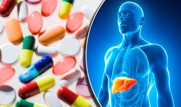
Drug-induced organ toxicity is a significant clinical challenge, representing a common cause of morbidity and mortality. While any organ system can be affected by drugs (including the kidneys, heart, lungs, skin, and bone marrow), the liver holds a unique position due to its central role in drug metabolism, detoxification, and excretion. Consequently, the liver is particularly vulnerable to drug-induced injury, leading to the complex and varied syndrome known as Drug-Induced Liver Injury (DILI). Recognizing, diagnosing, and managing DILI are crucial skills for healthcare professionals.
Drug-Induced Organ Reactions (Focus on the Liver)
Drugs exert their therapeutic effects by interacting with biological targets. However, these interactions, or the body’s processing of the drug, can sometimes lead to unintended adverse effects on various organs. The liver, being the primary site for biotransformation of most xenobiotics (foreign compounds, including drugs), is exposed to high concentrations of parent drugs, their metabolites, and potential toxins absorbed from the gut via the portal circulation.
Drug-induced liver injury can occur through various mechanisms:
- Intrinsic Toxicity: Predictable, dose-dependent injury inherent to the pharmacological properties of the drug or its metabolites. This type often has a short latency period and affects a significant proportion of exposed individuals (e.g., paracetamol overdose).
- Idiosyncratic Toxicity: Unpredictable, non-dose-dependent reactions that occur in a small subset of individuals. These are not related to the known pharmacological effects of the drug and are thought to involve host-specific factors (genetic predisposition, immune reactions, metabolic differences) and environmental triggers. Idiosyncratic DILI is less common but often more severe than intrinsic DILI.
DILI can present with a wide spectrum of severity, from asymptomatic laboratory abnormalities to acute liver failure requiring transplantation or resulting in death. It can also lead to chronic liver disease, such as chronic hepatitis or cirrhosis.
Classification of Common DILI Types
DILI is typically classified based on the pattern of liver enzyme abnormalities observed in blood tests, often reflecting the primary site and mechanism of injury within the liver parenchyma or biliary tree. The three main patterns are:
- Hepatocellular Injury:
- Definition: Characterized by a predominant elevation in serum aminotransferases (Alanine Aminotransferase (ALT) and Aspartate Aminotransferase (AST)), suggesting damage to hepatocytes. The ratio of ALT or AST to Alkaline Phosphatase (ALP) is typically high. A commonly used criterion is ALT elevation ≥ 5 times the upper limit of normal (ULN), or ALT ≥ 3 times ULN and ALP < 2 times ULN.
- Mechanism: Often related to direct hepatocyte damage, metabolic activation of the drug into toxic metabolites, immune-mediated reactions against hepatocytes, or interference with hepatocellular processes. Intrinsic DILI often presents with a hepatocellular pattern.
- Examples:
- Paracetamol (Acetaminophen): At high doses (intrinsic), causes severe centrilobular necrosis.
- Isoniazid: Can cause severe, sometimes fatal, hepatocellular injury, especially in slow acetylators (idiosyncratic, metabolic/immune mechanism).
- Statins (HMG-CoA reductase inhibitors): Usually cause mild, asymptomatic ALT elevation, but rarely can lead to severe acute hepatocellular injury (idiosyncratic).
- NSAIDs (Nonsteroidal Anti-inflammatory Drugs): Various mechanisms, can cause acute or chronic hepatitis (idiosyncratic).
- Antituberculosis drugs (e.g., Rifampicin, Pyrazinamide): Can cause significant hepatocellular damage.
- Cholestatic Injury:
- Definition: Characterized by a predominant elevation in serum Alkaline Phosphatase (ALP) and Gamma-Glutamyl Transferase (GGT), often accompanied by hyperbilirubinemia (jaundice), suggesting impaired bile flow. Aminotransferase levels may be normal or only mildly elevated. A commonly used criterion is ALP elevation ≥ 2 times ULN, or ALP ≥ 1.5 times ULN and ALT < 2 times ULN.
- Mechanism: Involves disruption of bile formation and/or flow, potentially due to damage to cholangiocytes (bile duct cells), disruption of bile acid transporters, canalicular damage, or immune-mediated attack on the biliary system.
- Examples:
- Amoxicillin-Clavulanate: A classic cause of delayed cholestatic or mixed injury, often presenting weeks after starting or stopping the drug (idiosyncratic, likely immune).
- Chlorpromazine (and other phenothiazines): Can cause prolonged cholestasis (idiosyncratic).
- Anabolic Steroids: Can cause dose-dependent intrahepatic cholestasis (intrinsic, related to transporter inhibition).
- Erythromycin Estolate: Historically known to cause cholestatic hepatitis.
- Mixed Injury:
- Definition: Characterized by significant elevation of both aminotransferases and alkaline phosphatase. The ratio of ALT/ALP falls between the criteria for hepatocellular and cholestatic injury. A commonly used criterion is ALT ≥ 3 times ULN and ALP ≥ 1.5 times ULN.
- Mechanism: Involves damage to both hepatocytes and the biliary system.
- Examples:
- Phenytoin: Can cause mixed or hepatocellular injury, sometimes part of a hypersensitivity reaction syndrome (idiosyncratic, immune/metabolic).
- Sulfonamides (e.g., Trimethoprim-Sulfamethoxazole): Can cause mixed injury, often with fever, rash, and eosinophilia (idiosyncratic, immune).
- Lisinopril (and other ACE inhibitors): Although rare, can cause mixed or cholestatic injury.
It’s important to note that some drugs can cause various patterns of injury, and the presentation can evolve over time.
Approach to the Patient with Presumed DILI (Step-by-Step Guide)
Diagnosing DILI is often a challenging process as it is primarily a diagnosis of exclusion. A systematic approach is crucial:
- Step 1: Maintain a High Index of Suspicion & Obtain a Detailed History:
- Consider DILI in any patient with new onset liver enzyme abnormalities or jaundice, especially if they are taking medications.
- Thoroughly review all medications, including prescription drugs, over-the-counter drugs, herbal supplements, dietary supplements, and recreational drugs. Obtain start and stop dates and dosages.
- Inquire about the timing of liver abnormalities relative to drug exposure. Latency can range from days to months or even years, and injury can occur even after stopping the drug (especially cholestatic).
- Ask about symptoms (fatigue, nausea, abdominal pain, dark urine, pale stools, jaundice, pruritus) and signs of hypersensitivity (rash, fever, eosinophilia, arthralgia).
- Assess alcohol consumption, travel history, exposure to viral hepatitis risks, and family history of liver disease.
- Step 2: Perform a Physical Examination:
- Look for signs of jaundice, hepatomegaly, splenomegaly, abdominal tenderness.
- Assess for signs of chronic liver disease or portal hypertension (ascites, edema, spider angiomata, palmar erythema).
- Check for signs of systemic hypersensitivity (rash, lymphadenopathy).
- Assess neurological status for signs of hepatic encephalopathy.
- Step 3: Conduct Initial Investigations:
- Liver Function Tests (LFTs): Measure ALT, AST, ALP, GGT, total and direct bilirubin, albumin, and International Normalized Ratio (INR). Determine the pattern (hepatocellular, cholestatic, or mixed).
- Complete Blood Count (CBC): Look for eosinophilia (suggesting hypersensitivity), anemia, or thrombocytopenia.
- Renal Function Tests (Creatinine, Urea, Electrolytes): Assess kidney involvement (potentially part of a systemic reaction or hepatorenal syndrome in advanced injury).
- Imaging: Abdominal ultrasound is often performed to evaluate liver size and echotexture, assess for signs of chronic liver disease, and rule out extrahepatic biliary obstruction (e.g., gallstones, mass) that could cause secondary liver injury or mimic cholestatic DILI.
- Step 4: Rule Out Other Causes of Liver Disease:
- This is a critical step, as DILI is a diagnosis of exclusion. Tests should include:
- Markers for acute viral hepatitis (Hepatitis A IgM, Hepatitis B surface antigen and core IgM antibody, Hepatitis C antibody).
- Testing for other less common infections (e.g., CMV, EBV, HSV) if clinically suspected.
- Autoimmune markers (ANA, ASMA, anti-LKM1, IgG levels) to rule out autoimmune hepatitis.
- Iron studies (ferritin, transferrin saturation) to rule out hemochromatosis.
- Alpha-1 Antitrypsin levels to rule out alpha-1 antitrypsin deficiency.
- Ceruloplasmin and serum copper to rule out Wilson’s disease (consider especially in younger patients).
- Specific tests based on history (e.g., celiac disease antibodies if malabsorption features).
- This is a critical step, as DILI is a diagnosis of exclusion. Tests should include:
- Step 5: Assess Causality:
- While challenging, formal causality assessment tools exist, such as the Roussel Uclaf Causality Assessment Method (RUCAM) or the Naranjo scale. These tools consider factors like the timing of onset relative to drug exposure, the clinical pattern of injury, exclusion of other causes, response to drug withdrawal, and prior knowledge of the drug’s hepatotoxicity.
- Step 6: Discontinue the Suspect Drug(s):
- If DILI is considered likely, the implicated drug(s) must be stopped immediately. This is the cornerstone of DILI management. Improvement in liver tests after drug withdrawal strongly supports the diagnosis.
- Step 7: Monitor and Provide Supportive Care:
- Monitor LFTs and clinical status regularly to assess improvement or worsening.
- Manage symptoms like pruritus (e.g., with cholestyramine) and nausea.
- Provide nutritional support.
- Monitor for complications of liver failure if severe injury occurs (coagulopathy, encephalopathy, renal impairment).
- Step 8: Consider Liver Biopsy:
- Liver biopsy is not always necessary but can be helpful in certain situations:
- To confirm the diagnosis if causality is uncertain or other diagnoses cannot be excluded.
- To assess the severity of injury and prognosis.
- To evaluate for superimposed or confounding liver diseases.
- To assess for features indicative of chronic DILI or progression to fibrosis/cirrhosis.
- Liver biopsy is not always necessary but can be helpful in certain situations:
Management of Some Important Cases of DILI: Paracetamol Overdose
Paracetamol (acetaminophen) overdose is the most common cause of intrinsic DILI globally and a leading cause of acute liver failure.
- Mechanism: At therapeutic doses, paracetamol is primarily metabolized in the liver by sulfation and glucuronidation into non-toxic conjugates. A small amount is metabolized by cytochrome P450 enzymes (mainly CYP2E1) into a highly reactive, toxic intermediate metabolite, N-acetyl-p-benzoquinone imine (NAPQI). NAPQI is rapidly detoxified by conjugation with glutathione. In overdose, the sulfation and glucuronidation pathways become saturated, leading to increased metabolism by CYP450, overwhelming glutathione stores. The excess NAPQI then binds covalently to hepatocellular proteins, causing oxidative stress and cell death.
- Clinical Course: Symptoms in the first 24 hours may be minimal (nausea, vomiting). Liver injury typically becomes evident 24-72 hours after ingestion, with rising aminotransferases, potentially progressing to jaundice, coagulopathy, encephalopathy, and multi-organ failure.
- Management:
- Risk Assessment: Determine the ingested dose, time since ingestion, and any co-ingestants (e.g., alcohol, other enzyme inducers).
- Laboratory Tests: Measure plasma paracetamol level (especially at 4 hours post-ingestion or later), LFTs, INR, renal function, and electrolytes.
- Decontamination: Activated charcoal may be administered if the patient presents within 1-2 hours of ingestion and the dose is significant.
- Antidote: The specific antidote is N-acetylcysteine (NAC). NAC acts by repleting glutathione stores, promoting NAPQI detoxification, and potentially having other protective effects. NAC is highly effective at preventing hepatotoxicity if administered within 8 hours of ingestion, but it offers benefits even when given later, particularly in established injury or acute liver failure. It is given intravenously or orally according to standard protocols (e.g., the 21-hour intravenous protocol).
- Supportive Care: Manage nausea, ensure hydration, correct electrolyte abnormalities, and monitor for signs of liver failure.
- Liver Transplant: Patients developing severe acute liver failure unresponsive to NAC may require evaluation for urgent liver transplantation based on prognostic criteria such as the King’s College Criteria.
Major Groups of Drugs to Avoid or Use with Extreme Caution in Liver Cirrhosis
Patients with liver cirrhosis have impaired liver function, reduced hepatic blood flow, portosystemic shunting, and often portal hypertension. These physiological changes significantly alter drug pharmacokinetics (absorption, distribution, metabolism, excretion) and pharmacodynamics, increasing the risk of adverse drug reactions and complications. Certain drug classes pose particular risks:
- Sedatives, Hypnotics, Opioids, and Psychoactive Drugs (e.g., Benzodiazepines, Narcotics, Antipsychotics): The liver clears many of these drugs. Impaired metabolism can lead to prolonged half-lives, accumulation, and increased sensitivity, significantly increasing the risk of precipitating or worsening hepatic encephalopathy.
- Nonsteroidal Anti-inflammatory Drugs (NSAIDs): Even short-term use can cause significant issues. NSAIDs inhibit renal prostaglandins, predisposing patients to impaired renal function, hepatorenal syndrome, and electrolyte disturbances. They also damage the gastric mucosa and platelet function, increasing the risk of peptic ulcers and gastrointestinal bleeding, which is particularly dangerous in the presence of esophageal or gastric varices.
- Diuretics (especially Loop Diuretics without Potassium-Sparing Agents): While diuretics are often necessary for ascites management, their use requires caution. Aggressive diuresis can lead to volume depletion, electrolyte imbalances (hyponatremia, hypokalemia), and precipitate hepatorenal syndrome or encephalopathy.
- Certain Antibiotics & Drugs with Significant Hepatic Metabolism or Hepatotoxicity: Drugs primarily metabolized by the liver may require dose reduction or avoidance. Some antibiotics (e.g., Trimethoprim-Sulfamethoxazole, Ciprofloxacin in some cases) can contribute to encephalopathy by altering gut flora and increasing ammonia production. Potentially hepatotoxic drugs should generally be avoided unless absolutely necessary and with close monitoring.
- Alcohol: Continued alcohol consumption in patients with cirrhosis is critical and significantly worsens liver disease and prognosis. It is mandatory to avoid alcohol entirely.
- Iron Supplements: Should be avoided unless there is a clear indication (e.g., iron deficiency anemia unrelated to variceal bleeding) and ideally after excluding hereditary hemochromatosis as the cause of cirrhosis, as iron overload can worsen damage.
- Beta-Blockers (Non-selective, e.g., Propranolol, Nadolol): While used to reduce portal pressure and prevent variceal bleeding, caution is needed, especially in advanced cirrhosis or in the presence of ascites, as they can worsen renal perfusion and precipitate hepatorenal syndrome or affect hemodynamics in ways that are detrimental. Dosage should be carefully titrated.
- Renin-Angiotensin System Inhibitors (ACE inhibitors, ARBs): Can exacerbate decreased renal perfusion in patients with ascites and predispose to hepatorenal syndrome.
It is imperative to review the medication list for every patient with cirrhosis regularly, adjusting doses, substituting safer alternatives, or discontinuing unnecessary drugs to minimize the risk of complications. Consultation with a pharmacist or hepatologist is often beneficial.
Conclusion
Drug-induced liver injury is a multifaceted and challenging clinical entity. A thorough understanding of its patterns, a systematic approach to diagnosis involving careful history taking and exclusion of other causes, and awareness of specific toxicity profiles (like paracetamol) are essential. Furthermore, recognizing the altered drug metabolism and increased risks in patients with underlying liver disease, particularly cirrhosis, is paramount to preventing further harm. Vigilance and careful medication management are key to optimizing outcomes in patients susceptible to or affected by DILI.


