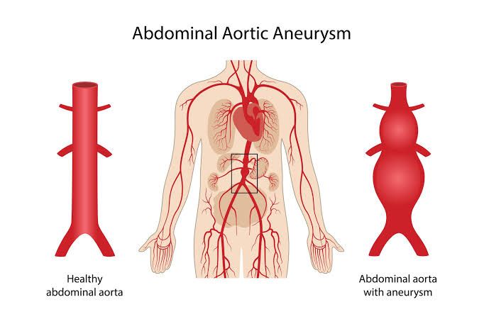
Arterial aneurysms represent localized dilatations or ballooning of an artery wall, resulting from weakening of the vessel’s structural components. While potentially present in any artery, they exhibit predilection for certain sites and carry varying risks depending on their location, size, and morphology.
Common Sites and Relative Incidence of Arterial Aneurysms
Arterial aneurysms can occur throughout the arterial system, but their prevalence varies significantly by site. The most common locations are:
- Aorta: The largest artery in the body, the aorta is the most frequent site for aneurysm formation.
- Abdominal Aortic Aneurysm (AAA): By far the most common type, AAAs typically occur below the level of the renal arteries. They are significantly more common in men than women and the incidence increases with age, peaking in individuals over 65. The estimated prevalence in men over 65 is around 4-8%.
- Thoracic Aortic Aneurysm (TAA): Occurring in the chest, TAAs are less common than AAAs but still represent a significant proportion of aortic aneurysms. They can involve the aortic root, ascending aorta, aortic arch, or descending aorta. The incidence is lower than AAAs, perhaps around 10-30% of all aortic aneurysms, but they pose unique challenges due to their proximity to vital structures and the dynamics of aortic flow.
- Peripheral Arteries: Aneurysms can also affect arteries in the limbs and other regions.
- Popliteal Artery Aneurysm: The most common peripheral artery aneurysm, located behind the knee. Popliteal aneurysms are often bilateral and frequently associated with AAAs; approximately 50% of patients with a popliteal aneurysm also have an AAA. They are much less common than aortic aneurysms, but carry a significant risk of thrombosis or embolization leading to limb ischemia.
- Femoral Artery Aneurysm: Occurs in the groin area. Less common than popliteal aneurysms, they are also sometimes associated with AAA.
- Visceral Artery Aneurysms: Aneurysms of arteries supplying abdominal organs (e.g., splenic, hepatic, renal, mesenteric arteries). Splenic artery aneurysms are the most common visceral type and are more frequent in women, especially multiparous women and those with portal hypertension.
- Cerebral Aneurysms: Aneurysms in the arteries of the brain, commonly found at arterial bifurcations in the Circle of Willis. While distinct in pathology and management, they represent another significant category of arterial aneurysm with a high risk of rupture (subarachnoid hemorrhage).
In summary of relative incidence, AAAs are the most prevalent, followed by TAAs, and then peripheral aneurysms (primarily popliteal), with cerebral and visceral aneurysms being less frequent depending on specific populations.
Rupturing Abdominal Aortic Aneurysm (AAA): Symptoms, Signs, Diagnosis, and Management
A ruptured AAA is a life-threatening surgical emergency requiring immediate recognition and intervention.
- Symptoms and Signs: The classic triad is:
- Severe abdominal or back pain: Often sudden onset, tearing or ripping in nature, and may radiate to the flank, groin, or legs.
- Hypotension or shock: Due to significant blood loss into the retroperitoneum or peritoneal cavity. Signs include dizziness, fainting, pallor, diaphoresis, rapid heart rate, and low blood pressure.
- Pulsatile abdominal mass: May be palpable, though its absence does not rule out a rupture, especially in obese patients or those in severe shock. Other potential signs include flank ecchymosis (Grey Turner’s sign) or periumbilical ecchymosis (Cullen’s sign), though these are often late findings. Some ruptures may be contained, presenting with pain but without overt shock initially, offering a brief window for intervention.
- Differential Diagnosis: The symptoms of a ruptured AAA can overlap with several other acute conditions, making accurate and rapid diagnosis crucial. Key differential diagnoses include:
- Renal colic (especially with severe flank pain)
- Acute pancreatitis
- Diverticulitis or colitis
- Myocardial infarction (especially with referred abdominal pain)
- Mesenteric ischemia or bowel obstruction
- Acute back pain (musculoskeletal or discogenic)
- Retroperitoneal hemorrhage from other causes
- Acute cholecystitis or biliary colic
- Diagnostic Plan: Diagnosis must be swift.
- Clinical Suspicion: A high index of suspicion based on the patient’s age, risk factors (smoking, hypertension), and presenting symptoms is paramount.
- Focused Bedside Ultrasound (FAST or dedicated AAA scan): Can rapidly identify the presence of an AAA and free fluid in the abdomen, highly suggestive of rupture. This is often the initial rapid test in unstable patients.
- Computed Tomography Angiography (CTA): The gold standard for diagnosis in hemodynamically stable patients. CTA confirms the presence of the AAA, identifies the rupture site, assesses the extent of hemorrhage, and provides detailed anatomical information crucial for surgical planning (open vs. endovascular repair). Unstable patients should proceed directly to the operating theater without extensive imaging if the diagnosis is strongly suspected clinically or confirmed by bedside ultrasound.
- Other Tests: Blood tests (complete blood count, electrolytes, renal function, coagulation profile, blood type and crossmatch) are essential for assessing the patient’s physiological status and preparing for transfusion.
- Management Plan: Management is focused on immediate resuscitation and definitive surgical repair.
- Immediate Call for Help: Activate the trauma team or vascular surgery service immediately.
- Resuscitation: Establish large-bore intravenous access. Administer intravenous fluids and blood products (packed red blood cells, fresh frozen plasma, platelets) to support blood pressure and organ perfusion. Aggressive blood pressure normalization should be avoided (“permissive hypotension”) initially, as it can dislodge clots and increase bleeding until surgical control is achieved.
- Pain Control: Administer analgesia.
- Monitoring: Continuous monitoring of vital signs, ECG, and urine output.
- Rapid Transport: Transfer the patient immediately to an operating room equipped for vascular surgery.
- Definitive Repair: Surgical intervention is mandatory. This can be:
- Open Surgical Repair: Involves laparotomy, clamping the aorta above and below the aneurysm, opening the aneurysm sac, and replacing the diseased segment with a synthetic graft. This is the traditional method and often necessary for unstable patients or unfavorable anatomy for EVAR.
- Endovascular Aneurysm Repair (EVAR): A less invasive technique involving placing a stent graft within the aneurysm through small incisions (typically in the groin). EVAR is preferred when anatomically feasible and the patient’s condition allows, as it is associated with lower perioperative morbidity and mortality compared to open repair in stable or contained ruptures.
Indications, Contraindications, and Risk Factors for Surgery in Chronic Asymptomatic Abdominal Aortic Aneurysms
Elective repair of asymptomatic AAAs is performed to prevent rupture, balancing the risk of rupture against the risks of the repair procedure.
- Indications for Elective Repair:
- Size: The primary indication. The generally accepted threshold is an maximal diameter of ≥ 5.5 cm in men and often ≥ 5.0 cm in women due to potentially higher rupture risk at smaller sizes.
- Rapid Expansion: An increase in diameter of ≥ 0.5 cm over a 6-month period or ≥ 1 cm per year is a strong indication, regardless of absolute size below the threshold, as rapid expansion predicts increased rupture risk.
- Symptoms: While discussed as asymptomatic, some patients report vague abdominal or back pain that is attributable to the aneurysm (e.g., pulsatile sensation, abdominal fullness). These symptoms, if clearly linked to the aneurysm, warrant repair.
- Saccular Shape: Saccular (pocket-like) aneurysms, even at smaller sizes, may have a higher rupture risk than fusiform (spindle-shaped) aneurysms and might warrant earlier intervention.
- Presence of Thrombus/Mural Irregularity: While less definitive, some morphological features may influence the decision, though size remains the dominant factor.
- Contraindications for Elective Repair: The primary contraindication is when the risks of surgery outweigh the potential benefit of preventing rupture during the patient’s expected remaining lifespan. This includes:
- Severe Comorbidities: Such as advanced heart failure, severe ischemic heart disease with uncorrectable angina, very severe chronic obstructive pulmonary disease (COPD), end-stage renal disease not on dialysis, or severe neurological deficits (e.g., recent stroke with significant disability).
- Limited Life Expectancy: Due to advanced age or severe underlying medical conditions (e.g., metastatic cancer).
- Patient Refusal: Competent patient declining the procedure.
- Unsuitable Anatomy: For both open and EVAR techniques in certain complex cases (though newer techniques are expanding treatment options). Severe hostile abdomen from prior surgery can be a relative contraindication to open repair.
- Risk Factors for Surgery: These are factors that increase the morbidity and mortality associated with aneurysm repair.
- Patient Factors:
- Aneurysm Factors:
- Suprarenal involvement (for open repair)
- Tortuosity or complex anatomy (for EVAR)
- Inflammatory aneurysms (for open repair)
- Procedural Factors:
- Type of Repair (Open repair generally carries higher perioperative risks than EVAR, though long-term outcomes can differ)
- Urgency of procedure (ruptured repair carries much higher risk than elective)
- Experience of the surgical team and center
Prevention of Common Complications Following Aneurysm Surgery
Aneurysm repair, whether open or endovascular, carries risks of significant complications. Prevention strategies are crucial for optimizing outcomes.
- Cardiovascular Complications (Myocardial Infarction, Arrythmias):
- Prevention: Thorough preoperative cardiac risk assessment and optimization (ECG, stress testing, possible coronary revascularization), aggressive medical management of hypertension and hyperlipidemia, beta-blockade, statin therapy, careful intraoperative monitoring and management of fluid balance and hemodynamics, aggressive postoperative cardiac monitoring.
- Pulmonary Complications (Pneumonia, Respiratory Failure):
- Prevention: Preoperative assessment of pulmonary function and optimization (smoking cessation, bronchodilators), careful anesthetic management, early mobilization postoperatively, incentive spirometry, aggressive pulmonary hygiene, pain control permitting deep breathing.
- Renal Complications (Acute Kidney Injury):
- Prevention: Preoperative assessment of renal function, adequate hydration, avoiding nephrotoxic agents (NSAIDs, certain antibiotics, contrast media used in angiography), careful use of aortic clamping (open repair) to minimize renal artery ischemia, maintaining adequate renal perfusion pressure during and after surgery.
- Neurological Complications (Stroke, Spinal Cord Ischemia):
- Prevention: Preoperative assessment of carotid disease, careful blood pressure management during and after surgery to maintain cerebral perfusion, minimizing cerebral emboli during aortic manipulation. Spinal cord ischemia (paraplegia/paraparesis) is a risk, particularly with open thoracoabdominal or extensive descending thoracic aortic repairs. Prevention involves maintaining spinal cord perfusion pressure (mean arterial pressure targets), preserving collateral flow, selective use of intercostal/lumbar artery reimplantation, and potentially cerebrospinal fluid (CSF) drainage.
- Graft Complications (Infection, Thrombosis, Pseudoaneurysm, Endoleak):
- Prevention: Strict aseptic technique during surgery, appropriate prophylactic antibiotics, careful handling and placement of the graft/stent graft, ensuring adequate outflow vessels, meticulous surgical technique to prevent suture line pseudoaneurysms (open repair). Endoleaks (persistent blood flow outside the stent graft but within the aneurysm sac) post-EVAR require careful planning, correct device sizing and deployment, and lifelong surveillance imaging to identify and manage.
- Limb Ischemia:
- Prevention: Careful technique to prevent embolization or thrombosis of peripheral arteries during clamping or manipulation, ensuring limb perfusion post-repair.
- Bowel Ischemia:
- Prevention: Particularly relevant in open repair of extensive or ruptured AAAs. Requires careful attention to preserving mesenteric artery flow, maintaining adequate systemic perfusion, and recognizing symptoms early.
Prevention is multifaceted, starting with comprehensive preoperative assessment and optimization, continuing with meticulous intraoperative technique, and concluding with vigilant postoperative monitoring and management.
Comparison of Thoracic, Abdominal, Femoral, and Popliteal Aneurysms
These aneurysms differ significantly in their typical presentation, complication profiles, and treatment approaches.
| Feature | Abdominal Aortic (AAA) | Thoracic Aortic (TAA) | Femoral Artery | Popliteal Artery |
|---|---|---|---|---|
| Presentation | Usually asymptomatic; discovered incidentally or surveillance. Symptomatic: vague back/abd pain. Rupture: severe pain, shock, pulsatile mass. | Often asymptomatic; discovered incidentally. Symptomatic: chest/back pain, cough, hoarseness, dyspnea (compression). Rupture: severe chest/back pain, shock. | Usually asymptomatic mass in groin. May cause pain, nerve compression. Risk of thrombosis/embolization. | Usually asymptomatic mass behind knee. High risk of thrombosis/embolization leading to limb ischemia (“trash foot”). Less common to rupture. |
| Complications | ||||
| Frequency of Rupture | HIGH (most common major complication) if size > threshold | Significant, but risk varies greatly by location (ascending higher risk than descending per unit size), size, and associated dissection. | Low | Very Low |
| Frequency of Dissection | RARE (true dissection). Aortic Dissection is a separate pathology primarily affecting the aorta. | MORE COMMON than in AAA, especially in ascending aorta (Type A) or descending aorta (Type B) as the cause or complication of the aneurysm/aortopathy. | RARE | RARE |
| Frequency of Thrombosis | Relatively Low (large lumen). Mural thrombus common within sac but rarely occlusive. | Relatively Low (large lumen). | Moderate | HIGH (most common complication leading to acute limb ischemia) |
| Frequency of Embolization | Low (though mural thrombus can embolize distally, less frequent than peripheral source). | Low. | Moderate (distal leg/foot emboli). | HIGH (leading to “trash foot” – critical limb ischemia) |
| Treatment | Surveillance if small. Elective repair (EVAR or Open) if size > threshold or symptomatic. Ruptured: Urgent EVAR or Open repair. | Surveillance if small. Elective repair (TEVAR or Open) if size > threshold, symptomatic, or expanding. Ruptured: Urgent repair. | Surveillance if small. Repair (Open or Endovascular) if size > threshold, symptomatic, or thrombosed/embolizing. | Repair (Open or Endovascular) usually indicated due to high risk of thrombosis/embolization, even if small, in good surgical candidates. |
Conclusion
Arterial aneurysms are a significant vascular pathology with varied presentations and risks depending on their location. Abdominal aortic aneurysms are the most common, posing a critical risk of rupture, particularly when symptomatic or large. Management strategies range from surveillance for smaller, asymptomatic aneurysms to urgent surgical intervention for rupture. Elective repair of appropriately sized aneurysms is aimed at preventing catastrophic rupture, but must be weighed against the significant surgical risks, which are mitigated by thorough preoperative assessment and meticulous complication prevention strategies. Peripheral aneurysms, while less prone to rupture, carry a higher risk of thrombotic and embolic complications leading to limb ischemia, necessitating different management considerations. Understanding the unique characteristics of aneurysms at different sites is crucial for accurate diagnosis, timely intervention, and optimal patient outcomes.


