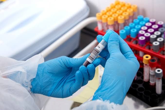
Cardiovascular diseases (CVDs) remain a leading cause of morbidity and mortality worldwide. Accurate and timely diagnosis is paramount for initiating appropriate treatment and improving patient outcomes. While clinical assessment, electrocardiography (ECG), and imaging techniques are fundamental diagnostic tools, biochemical markers, measured in blood, provide crucial objective evidence of myocardial injury, cardiac stress, or thrombosis. These biomarkers offer insights into cellular damage, physiological strain, and underlying pathological processes within the cardiovascular system.
The landscape of cardiac biomarkers has evolved significantly over time. Initially, relatively non-specific enzymes were utilized, providing evidence of tissue damage but lacking the specificity required for definitive cardiac diagnosis in many cases. More recently, highly sensitive and specific markers have become the cornerstone of diagnosing acute myocardial infarction (AMI) and other cardiac conditions. This discussion explores the role of both classic and contemporary biomarkers in the diagnosis and management of cardiovascular disease, structured to highlight the distinct contribution of each marker or group of markers.
Classic Cardiac Enzymes in the Diagnosis of Heart Disease: CK, LDH, and AST
Historically, several enzymes released from damaged tissue were used to detect myocardial injury. While largely superseded by more specific markers for acute coronary syndromes (ACS), understanding their role provides valuable context for the evolution of diagnostic strategies. The primary limitation of these enzymes is their widespread presence in various tissues, leading to poor specificity for cardiac damage when elevated.
- Creatine Kinase (CK) and its Isoenzymes:
- What it is: Creatine kinase is an enzyme found in tissues with high energy requirements, such as skeletal muscle, brain, and heart.
- Role in Diagnosis: Total CK elevation indicates muscle damage, but not specifically cardiac muscle. To improve specificity, isoenzymes are measured. The three main isoenzymes are:
- CK-BB (CK-1): Primarily found in the brain and nervous tissue.
- CK-MB (CK-2): Found predominantly in cardiac muscle (comprising about 15-40% of total CK in the myocardium) and in smaller amounts in skeletal muscle. This isoenzyme was historically the most specific enzyme marker for myocardial injury.
- CK-MM (CK-3): The major isoenzyme in skeletal muscle.
- Kinetic Profile in AMI: CK-MB typically begins to rise 4-6 hours after the onset of myocardial infarction, peaks around 24 hours, and returns to normal within 48-72 hours. Serial measurements showing a characteristic rise and fall were essential for diagnosis.
- Limitations: CK-MB is not entirely specific to cardiac muscle. Significant skeletal muscle injury (trauma, surgery, strenuous exercise, intramuscular injections) can also elevate CK-MB levels, albeit often without the characteristic rise and fall pattern seen in AMI. Its relatively late rise compared to newer markers is also a disadvantage in early diagnosis. Due to these limitations and the higher specificity and sensitivity of troponins, CK-MB is now less commonly used for routine AMI diagnosis.
- Lactate Dehydrogenase (LDH) and its Isoenzymes:
- What it is: Lactate dehydrogenase is an enzyme found in almost all body tissues, playing a role in anaerobic metabolism.
- Role in Diagnosis: Total LDH is a very non-specific marker of tissue damage. It exists as five isoenzymes (LDH-1 to LDH-5). LDH-1 is the predominant isoenzyme in cardiac muscle and red blood cells. In healthy individuals, LDH-2 levels are higher than LDH-1. Following myocardial injury, LDH-1 is released, often leading to a “flipped” ratio where LDH-1 > LDH-2.
- Kinetic Profile in AMI: LDH rises relatively late after MI, typically 10-12 hours after symptom onset, peaks between 48-72 hours, and remains elevated for 8-14 days. This prolonged elevation made it useful for detecting MI several days old, when CK-MB may have returned to normal.
- Limitations: LDH is released from numerous tissues (kidney, liver, skeletal muscle, red blood cells). Hemolysis (breakdown of red blood cells) is a common cause of elevated LDH-1 that can mimic or obscure cardiac injury. Its very late rise makes it unsuitable for early diagnosis or guiding acute management decisions. Consequently, LDH and its isoenzymes are rarely used in the modern diagnosis of AMI.
- Aspartate Aminotransferase (AST):
- What it is: Aspartate aminotransferase (also known as SGOT) is an enzyme involved in amino acid metabolism, found in high concentrations in the liver, heart, skeletal muscle, kidney, brain, and red blood cells.
- Role in Diagnosis: AST was one of the earliest enzymes used to diagnose MI.
- Kinetic Profile in AMI: AST typically rises 6-10 hours after symptom onset, peaks at 24 hours, and returns to normal within 4-7 days. It often paralleled the rise of CK.
- Limitations: AST has extremely poor specificity for cardiac injury due to its presence in so many tissues, particularly the liver. Elevated AST is more commonly associated with liver disease. Like LDH, its use in modern cardiac diagnostics is negligible.
In summary, while CK-MB, LDH, and AST provided initial biochemical evidence of cardiac damage, their lack of specificity, varying kinetics, and the advent of superior markers have rendered them largely obsolete for the routine diagnosis of acute myocardial injury.
Newer and More Specific Biomarkers in the Diagnosis of Cardiovascular Disease: Myoglobin, Troponins, Natriuretic Peptides, and D-dimers
Modern cardiac diagnostics relies heavily on biomarkers that offer higher sensitivity, specificity, and prognostic value.
- Myoglobin:
- What it is: Myoglobin is a small, oxygen-binding protein found in both cardiac and skeletal muscle.
- Role in Diagnosis: Myoglobin is released very rapidly from damaged muscle cells into the bloodstream.
- Kinetic Profile in suspected MI: Myoglobin is one of the earliest markers to rise, often detectable in blood within 1-4 hours of symptom onset. It peaks quickly (around 6-7 hours) and returns to normal within 24 hours due to renal clearance.
- Limitations: Myoglobin’s major limitation is its poor specificity for cardiac muscle. Any injury to skeletal muscle (trauma, strenuous exercise, kidney failure affecting clearance) will also elevate myoglobin levels.
- Clinical Utility: Despite its lack of specificity, myoglobin’s rapid release makes it useful as an early rule-out marker. A normal myoglobin level within a few hours of chest pain onset significantly reduces the likelihood of AMI at that time point. However, a positive myoglobin requires confirmation with more specific markers like troponin.
- Cardiac Troponins (cTnI and cTnT):
- What they are: Troponins are a complex of three proteins (Troponin I, Troponin T, and Troponin C) that regulate the interaction between actin and myosin in muscle contraction. Cardiac isoforms of Troponin I (cTnI) and Troponin T (cTnT) are found almost exclusively in cardiac muscle cells.
- Role in Diagnosis: Cardiac troponins are the gold standard biomarkers for detecting myocardial necrosis (heart muscle cell death). Even microscopic areas of damage release detectable amounts of these proteins.
- Kinetic Profile in AMI: Cardiac troponin levels typically begin to rise 3-6 hours after symptom onset (though high-sensitivity assays can detect elevations even earlier, within 1-3 hours), peak around 12-24 hours, and remain elevated for several days (often 7-14 days, sometimes longer). This prolonged elevation is a key advantage for diagnosis, especially if patients present late.
- Specificity and Sensitivity: Modern assays, particularly high-sensitivity cardiac troponin (hs-cTn) assays, are highly sensitive and specific for myocardial injury. Elevated cTn levels indicate cardiac muscle damage, but it’s crucial to understand that this damage can be caused by conditions other than acute myocardial infarction (Type 1 AMI, caused by plaque rupture and thrombosis). Other causes of elevated troponin include:
- Type 2 AMI (myocardial injury due to supply/demand mismatch, e.g., severe sepsis, tachycardia, severe anemia).
- Myocarditis (inflammation of the heart muscle).
- Pericarditis (inflammation of the sac surrounding the heart), especially with associated myocarditis.
- Pulmonary embolism.
- Acute heart failure.
- Chronic kidney disease.
- Severe hypertension.
- Tachyarrhythmias or bradyarrhythmias.
- Direct trauma to the heart (contusion).
- Clinical Utility: Cardiac troponins are central to the definition and diagnosis of myocardial infarction according to universal guidelines. Serial measurements are used to detect a significant rise and/or fall in levels, indicating acute injury. They are also valuable risk stratifiers in patients with ACS, with higher levels correlating with worse prognosis. hs-cTn assays allow for faster rule-in/rule-out protocols (e.g., 0/1h or 0/2h algorithms) in emergency settings, significantly improving diagnostic efficiency.
- Natriuretic Peptides (BNP and NT-proBNP):
- What they are: B-type natriuretic peptide (BNP) and its inactive precursor fragment, N-terminal pro-B-type natriuretic peptide (NT-proBNP), are hormones primarily released by the ventricles of the heart in response to increased wall stress and volume expansion.
- Role in Diagnosis: Natriuretic peptides are not markers of myocardial necrosis. Their primary role is in the diagnosis and management of heart failure (HF). Elevated levels indicate increased cardiac stretch and volume overload.
- Kinetic Profile: Levels rise in response to ventricular stretch and fall as heart failure improves with treatment.
- Clinical Utility: Elevated BNP or NT-proBNP levels in a patient presenting with dyspnea (shortness of breath) strongly suggest that the symptom is due to heart failure. They are used to differentiate heart failure from non-cardiac causes of dyspnea (e.g., lung disease). They also have significant prognostic value in patients with heart failure, with higher levels indicating more severe disease and increased risk of future events. BNP and NT-proBNP are also used to monitor the effectiveness of heart failure treatment.
- Limitations: Levels can be influenced by factors other than heart failure, such as age (increase with age), renal dysfunction (increase), obesity (decrease), and certain medications. They are also often elevated in acute coronary syndromes, not as a marker of necrosis, but due to accompanying ventricular dysfunction or stress.
- D-dimer:
- What it is: D-dimer is a fibrin degradation product, a small protein fragment produced when a blood clot dissolves in the body via fibrinolysis. Its presence indicates that clotting (thrombin activity) and clot breakdown (plasmin activity) are occurring somewhere in the body.
- Role in Diagnosis: D-dimer is not a direct marker of cardiac muscle damage or stress. Its role in cardiovascular disease is primarily related to assessing the presence of thrombosis.
- Clinical Utility: D-dimer is most commonly used to help rule out acute thromboembolic conditions, particularly pulmonary embolism (PE) and deep vein thrombosis (DVT), which can sometimes present with chest pain or shortness of breath that mimics cardiac symptoms or can occur concurrently with cardiac events. A normal D-dimer level in a patient with a low to intermediate clinical probability of PE or DVT effectively rules out these conditions.
- Limitations: D-dimer is elevated in many conditions besides PE/DVT, including recent surgery or trauma, infection, inflammation, cancer, pregnancy, liver disease, and disseminated intravascular coagulation (DIC). It is also often elevated in acute coronary syndromes, not as a cause, but as a consequence of systemic inflammatory and prothrombotic states. Therefore, an elevated D-dimer is highly non-specific and indicates only that clotting and fibrinolysis are occurring; it does not pinpoint the location or cause. Its value lies primarily in its high negative predictive value.
Integration and Clinical Context
It is critical to emphasize that biomarkers are only one piece of the diagnostic puzzle. Results must always be interpreted within the full clinical context, including the patient’s medical history, physical examination findings, ECG results, and imaging studies. Serial measurements of markers like cardiac troponins are often necessary to assess the dynamic changes characteristic of acute events. The specific marker used, the assay sensitivity, and local protocols can influence the diagnostic approach.
Conclusion
The use of cardiac and related biomarkers has revolutionized the diagnosis and management of cardiovascular diseases. While classic enzymes like CK, LDH, and AST provided early insights, they have been largely replaced by more specific and sensitive markers. Cardiac troponins (cTnI and cTnT) are the definitive biomarkers for diagnosing myocardial necrosis, central to the identification of acute myocardial infarction. Myoglobin offers potential for early rule-out but lacks specificity. Natriuretic peptides (BNP and NT-proBNP) are invaluable for the diagnosis and management of heart failure. D-dimer serves a distinct role in ruling out thromboembolic events. As diagnostic tools, these biomarkers provide objective evidence that, when combined with clinical assessment, ECG, and imaging, enables accurate diagnosis, risk stratification, and guide appropriate therapeutic interventions, ultimately improving patient care in the complex landscape of cardiovascular disease.


