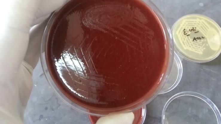
Accurate diagnosis of sexually transmitted diseases (STDs), also known as sexually transmitted infections (STIs), is paramount for effective patient management, disease control, and public health. The reliability of diagnostic testing is heavily dependent on the quality of the specimen collected, its proper storage and transport, and the correct application of laboratory techniques such as microscopy and culture.
Specimen Collection Methods for Sexually Transmitted Diseases
The appropriate method of specimen collection varies depending on the suspected pathogen and the site of infection. Strict adherence to aseptic technique and specific site protocols is essential to obtain a representative sample while minimizing contamination.
General Principles for Specimen Collection:
- Patient Identification: Confirm patient identity using at least two identifiers.
- Patient Preparation: Provide clear instructions to the patient (e.g., avoid urinating for a specified time before collecting urethral or first-catch urine specimens, avoid douching before cervical samples).
- Proper Supplies: Use sterile collection devices and the correct transport media specified by the laboratory.
- Aseptic Technique: Maintain sterility during collection to prevent contamination.
- Labeling: Label specimens accurately and immediately after collection with patient name, date of birth, collection date and time, and specimen source.
- Safety: Use standard precautions (gloves, protective eyewear) when handling potentially infectious materials.
Specific Specimen Collection Methods:
- Urethral Swab (primarily for males):
- Purpose: Detection of Neisseria gonorrhoeae, Chlamydia trachomatis, and sometimes Trichomonas vaginalis.
- Procedure:
- Instruct the patient not to urinate for at least 1-2 hours prior to collection.
- Use a sterile synthetic swab (not cotton, as it can contain inhibitors) on a plastic or wire shaft.
- Insert the swab 2-4 cm into the urethral meatus.
- Gently rotate the swab for 5-10 seconds to collect epithelial cells and exudate.
- Carefully withdraw the swab without touching the external genitalia.
- Immediately place the swab into the appropriate transport medium (e.g., transport medium for Neisseria culture, nucleic acid amplification test (NAAT) medium).
- First-Catch Urine (for both males and females):
- Purpose: Primarily for detection of Chlamydia trachomatis and Neisseria gonorrhoeae using NAATs. This is less invasive and often preferred.
- Procedure:
- Instruct the patient not to clean the genital area prior to collection.
- Instruct the patient to collect the first portion (20-30 mL) of the voided urine stream into a sterile collection cup. Do not collect midstream or a large volume sample, as pathogens are most concentrated in the first void.
- Transfer the urine to the specific transport tube provided for NAAT testing according to the manufacturer’s instructions.
- Cervical Swab (for females):
- Purpose: Detection of Neisseria gonorrhoeae, Chlamydia trachomatis, and potentially Trichomonas vaginalis.
- Procedure:
- Perform a speculum examination to visualize the cervix.
- Remove excess cervical mucus using a separate swab and discard it. This step is crucial as mucus can interfere with testing.
- Insert a sterile synthetic swab into the endocervical canal (1-2 cm).
- Rotate the swab gently but firmly against the cervical wall for 10-30 seconds to collect columnar epithelial cells.
- Withdraw the swab carefully.
- Place the swab into the appropriate transport medium (e.g., transport medium for Neisseria culture or NAAT medium).
- Rectal Swab:
- Purpose: Detection of Neisseria gonorrhoeae and Chlamydia trachomatis in symptomatic or asymptomatic individuals.
- Procedure:
- Insert a sterile synthetic swab 2-4 cm into the anal canal.
- Rotate the swab gently against the rectal wall for 10-30 seconds.
- Carefully withdraw the swab, avoiding contamination with gross fecal material.
- Place the swab into the appropriate transport medium.
- Pharyngeal Swab:
- Purpose: Detection of Neisseria gonorrhoeae and Chlamydia trachomatis in the pharynx.
- Procedure:
- Instruct the patient to open their mouth and say “ahh”.
- Using a tongue depressor, carefully swab the posterior pharynx and tonsillar areas, avoiding the tongue and uvula.
- Place the swab into the appropriate transport medium.
- Lesion Swab:
- Purpose: Detection of Herpes Simplex Virus (HSV) or Treponema pallidum (syphilis).
- Procedure:
- Clean the lesion surface with sterile saline or water to remove crust and debris.
- For HSV: Swab the base of a fresh vesicle or early ulcer with a viral transport swab. Break open a vesicle if necessary to collect fluid. Place the swab in Viral Transport Medium (VTM).
- For Treponema pallidum (Darkfield Microscopy): Gently abrade the lesion base to produce serous fluid. Collect the fluid onto a glass slide or into a capillary tube. This procedure requires immediate examination.
Specimen Storage and Transport
Proper storage and timely transport are critical for maintaining the viability of organisms (for culture) and preserving the integrity of nucleic acids or antigens (for other tests like NAATs).
- General Rule: Process specimens as quickly as possible after collection.
- Refrigeration (2-8°C): Most bacterial specimens (e.g., swabs in Amies or Stuart transport media), viral transport media (VTM), and urine for NAAT testing can be refrigerated if processing is delayed. Exception: Neisseria gonorrhoeae is sensitive to cold and drying.
- Room Temperature: Specimens for Neisseria gonorrhoeae culture should be transported at room temperature using specific transport systems (e.g., JEMBEC system with a CO2-generating tablet, Amies with charcoal swab in a robust system). Specimens for Trichomonas vaginalis wet mount should be kept at room temperature and examined immediately.
- Specific Transport Media: Always use the transport medium provided by the testing laboratory, as it is optimized for preserving specific pathogens and is compatible with their testing methods.
- Packaging: Package specimens securely in leak-proof containers and follow biohazard shipping regulations if transporting off-site. Include the completed requisition form, placed outside the primary specimen container to prevent contamination.
Microscopic Recognition of Pathogens in Urethral Discharge
Microscopy, particularly Gram stain and wet mount, is a rapid technique for examining urethral discharge and can provide immediate clues for diagnosis and initial treatment.
A. Gram Stain:
- Preparation:
- Place a small amount of urethral discharge onto a clean glass slide.
- Spread thinly and evenly.
- Allow to air dry completely.
- Heat fix the slide gently by passing it through a flame quickly, or use methanol fixation (cover with methanol for 1 minute, drain, air dry) which is often preferred for preserving cell morphology.
- Staining Procedure: Perform standard Gram staining steps:
- Flood slide with Crystal Violet (primary stain) for 1 minute. Rinse gently with water.
- Flood slide with Gram’s Iodine (mordant) for 1 minute. Rinse gently with water.
- Decolorize briefly with Gram’s decolorizer (alcohol-acetone mixture) until the solvent runs clear (usually a few seconds). This step is critical and requires careful timing. Rinse immediately with water.
- Flood slide with Safranin (counterstain) for 30 seconds to 1 minute. Rinse gently with water.
- Blot dry gently or air dry.
- Microscopic Examination: Examine under a microscope, typically starting with lower power (10x) to find areas with discharge and cells, then 40x, and finally 100x (oil immersion) for detailed bacterial morphology.
- Interpretation (Key findings in urethral discharge):
- Neutrophils (Polymorphonuclear Leukocytes – PMNs): Look for the presence and quantity of WBCs. A high number of PMNs (e.g., >5-10 per high-power field (HPF) or >2-4 per oil immersion field (OIF) in specific areas) indicates inflammation and is suggestive of urethritis.
- Neisseria gonorrhoeae: Classically appears as Gram-negative diplococci (paired cocci), often coffee-bean or kidney-bean shaped. The most characteristic finding is the presence of intracellular Gram-negative diplococci within neutrophils. Extracellular Gram-negative diplococci should also be noted but are less specific.
- Other Bacteria: Observe for other bacterial morphologies (Gram-positive cocci in clusters or chains, Gram-negative rods, etc.). While not primary causes of gonococcal/chlamydial urethritis, their presence can indicate other infections or colonizing flora.
B. Wet Mount:
- Preparation:
- Place a drop of sterile physiological saline on a clean glass slide.
- Mix a small amount of urethral discharge into the saline using a swab or loop.
- Place a coverslip over the mixture, avoiding air bubbles.
- Microscopic Examination: Examine immediately at 10x and 40x magnification. Delay reduces the chance of observing organism motility.
- Interpretation (Key findings in urethral discharge):
- White Blood Cells (Leukocytes): Note their presence (similar to Gram stain, indicates inflammation).
- Epithelial Cells: Observe squamous or columnar epithelial cells.
- Trichomonas vaginalis: Look for motile organisms. They are pear-shaped with visible flagella and a characteristic jerky, tumbling, or wobbling motility. They are roughly the size of a large white blood cell. Motility is key to identification in a wet mount.
- Candida sp. (Yeast): May be seen as budding yeast cells (oval shapes) and possibly pseudohyphae (elongated chains of yeast cells). While typically associated with vaginal candidiasis, yeast balanitis (penile candidiasis) can occur and may be seen in urethral samples, or yeast may be present as part of normal flora.
Culture and Identification of Neisseria sp. (Simulated Specimen Example)
Culture remains important for identifying Neisseria gonorrhoeae, especially for antimicrobial susceptibility testing and confirming ambiguous NAAT results. This section details the process using a simulated specimen containing Neisseria sp.
A. Culture of Neisseria sp.
- Specimen Type: Simulated urethral discharge (e.g., broth or swab with added Neisseria sp.).
- Media: Use selective media designed for Neisseria gonorrhoeae, such as Thayer-Martin (TM), Modified Thayer-Martin (MTM), Martin-Lewis (ML), or NYC medium. These media contain antimicrobial agents (e.g., vancomycin, colistin, nystatin, trimethoprim) to suppress the growth of commensal bacteria and fungi while allowing Neisseria to grow.
- Procedure:
- Obtain a sterile selective agar plate.
- Using a sterile loop or the collection swab from the simulated specimen, inoculate the plate by spreading the material over a small area at one edge.
- Using a new sterile loop (or sterilizing the previous one), streak the inoculum from the initial area across the plate using the four-quadrant method or a continuous streaking pattern to obtain isolated colonies.
- Incubate the plate in a humid atmosphere enriched with 3-10% CO2 at 35-37°C for 24-48 hours. Neisseria gonorrhoeae is fastidious and requires these specific conditions for growth. Candle jars or CO2 incubators are commonly used.
B. Identification of Neisseria sp. from Culture
After incubation, examine the plates for characteristic colony morphology. N. gonorrhoeae typically forms small (0.5-1 mm), grey, translucent-to-opaque, raised colonies after 24-48 hours. Once potential Neisseria colonies are identified, perform the following steps:
- Gram Stain: Prepare a Gram stain from a suspected colony. Observe for Gram-negative diplococci. This confirms the organism is a Neisseria-like organism.
- Oxidase Test: All Neisseria species are oxidase positive. This is a rapid test that helps differentiate Neisseria from many other Gram-negative bacteria.
- Procedure: Place a piece of filter paper saturated with oxidase reagent on a clean surface. Using a sterile wooden or platinum loop (avoid nichrome loops which can give false positives), transfer a small amount of suspect colony onto the paper.
- Result: A positive test is indicated by the development of a purple or dark blue color within 10-30 seconds.
- Biochemical Identification (Carbohydrate Utilization Tests): These tests differentiate Neisseria species based on their ability to produce acid from specific carbohydrates.
- Principle: Organisms are inoculated into media containing specific sugars (glucose, maltose, lactose, sucrose) and a pH indicator. Acid production (due to fermentation or aerobic utilization) is indicated by a color change of the indicator.
- Common Methods:
- Cystine Tryptic Agar (CTA) Sugars: Tubes containing CTA base with individual sugars (glucose, maltose, lactose, sucrose). Inoculate heavily with organism and overlay with mineral oil to promote anaerobic metabolism within the agar column. Observe for yellow color change (acid production).
- Rapid Carbohydrate Utilization Tests (e.g., kits): Faster methods using pre-filled wells or strips with lyophilized substrates. Inoculate with a heavy suspension of the organism and incubate for a shorter time (e.g., 4 hours).
- Interpretation: The pattern of sugar utilization identifies the species:
- Neisseria gonorrhoeae: Acid production from Glucose ONLY.
- Neisseria meningitidis: Acid production from Glucose and Maltose.
- Neisseria lactamica: Acid production from Glucose, Maltose, and Lactose.
- Neisseria sicca/subflava group: Acid production from Glucose, Maltose, and Sucrose.
- Other Neisseria species (commensals): Various patterns, may utilize sucrose or none of the sugars tested.
- Confirmation: The combination of Gram stain appearance (Gram-negative diplococci), positive oxidase test, and the characteristic sugar utilization pattern (Glucose ONLY) confirms the isolation and identification of Neisseria gonorrhoeae.


