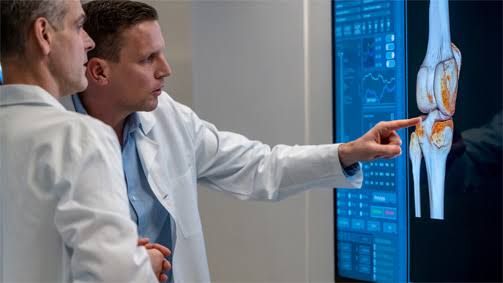
Accurate interpretation of musculoskeletal imaging studies is a cornerstone of patient care in numerous medical disciplines. It requires a solid foundation in anatomy, a methodical approach to identifying and characterizing pathology, and an understanding of the typical manifestations of various diseases across different imaging modalities.
Rapid Review of Essential Musculoskeletal Anatomy for Imaging
A detailed understanding of anatomy is paramount for interpreting musculoskeletal images. Location, relationships, and normal variants are fundamental to identifying pathology. This section outlines a rapid, structured approach focusing on anatomical points crucial for imaging interpretation. Think of this not as an exhaustive list, but as key “lines” or facts to recall quickly when reviewing an image.
Approach to Anatomical Review on Imaging:
When viewing any musculoskeletal image (X-ray, CT, MRI), adopt a systematic approach:
- Identify the Bone(s): Name the bones present. Familiarize yourself with the typical shape and size of the bones in that region.
- Assess Bone Components: Quickly review the major parts of the bone:
- Diaphysis: The shaft.
- Metaphysis: The wider part near the joint, adjacent to the physis (growth plate).
- Epiphysis: The end of the bone, articulating with other bones, contains the articular cartilage.
- Physis (Growth Plate): Present in skeletally immature individuals; a cartilaginous layer appearing lucent on X-ray. Crucial for assessing growth disturbances and fractures.
- Cortex: The dense outer layer. Assess its thickness and integrity.
- Medulla (Medullary Cavity): The inner spongy bone containing marrow. Assess its trabecular pattern.
- Periosteum: The outer membrane covering the bone (usually not visible on plain X-ray unless elevated by pathology).
- Evaluate the Joint(s): Identify the articular surfaces.
- Articular Cartilage: Covers the ends of bones in synovial joints (visible on MRI as intermediate signal intensity, lucent on X-ray – joint space). Assess its thickness and surface.
- Joint Space: Represents the thickness of the articular cartilage and meniscus/disc.
- Synovium: The lining of the joint capsule (visible on MRI, enhanced with contrast). Assess its thickness and pattern.
- Joint Capsule and Ligaments: Fibrous structures providing stability (best seen on MRI). Follow their course and assess integrity.
- Menisci/Discs: Fibrocartilaginous structures in certain joints (knee, wrist, spine, TMJ) providing cushioning and stability (best seen on MRI). Assess their shape and signal intensity.
- Survey Surrounding Soft Tissues:
- Muscles: Identify major muscle groups. Assess size, signal intensity (on MRI), and look for effusions/edema.
- Tendons: Connect muscle to bone. Identify major tendons in the region. Assess their course, caliber, and signal intensity.
- Bursae: Fluid-filled sacs reducing friction (often visible when inflamed/distended).
- Neurovascular Bundles: Be aware of the general location of major nerves and vessels, especially in cases of trauma or masses.
- Note Key Landmarks and Relationships: For specific regions (e.g., spine, shoulder, knee), know critical anatomical variants, common points of impingement, attachment sites, and typical fracture patterns associated with specific structures.
Examples of “Short Lines” (High-Yield Facts for Rapid Recall):
- Knee: Medial meniscus is C-shaped, lateral is O-shaped. ACL prevents anterior tibial translation, PCL prevents posterior. Patellar tendon connects patella to tibia; quadriceps tendon connects quadriceps muscle to patella.
- Shoulder: Rotator cuff muscles (SITS): Supraspinatus (most commonly torn), Infraspinatus, Teres Minor, Subscapularis. Biceps tendon (long head) runs through the bicipital groove. Glenoid labrum increases glenoid depth.
- Spine: Know the components of a vertebral segment (body, pedicles, laminae, spinous process, transverse processes, facets). Intervertebral disc anatomy (nucleus pulposus, annulus fibrosus). Know the spinal canal and foraminal boundaries.
- Wrist: Carpal bones in order (proximal row: Scaphoid, Lunate, Triquetrum, Pisiform; distal row: Trapezium, Trapezoid, Capitate, Hamate). Scaphoid is most commonly fractured carpal bone. Carpal tunnel contents (Median nerve and 9 tendons).
Regular review of normal anatomy on various imaging modalities is crucial. Comparing the image to known normal anatomy is the first step in identifying potential pathology.
Imaging Features of Benign and Malignant Bone Lesions
Characterizing bone lesions on imaging is critical to guide diagnosis and management. While biopsy is often necessary for definitive diagnosis, imaging features provide vital clues to narrow the differential diagnosis and assess the likelihood of benignity or malignancy.
Key Imaging Features to Evaluate:
When assessing a bone lesion, systematically evaluate the following features on plain radiographs, CT, and MRI:
- Location:
- Which bone?
- Which part of the bone (Epiphysis, Metaphysis, Diaphysis, Apophysis)?
- Eccentric or central within the medullary cavity?
- Cortical or medullary?
- Subchondral? Subperiosteal?
- Size and Shape:
- Measure the lesion.
- Is it round, oval, irregular?
- Margins (Zone of Transition): This is one of the most important features for differentiating benign from malignant.
- Sharp, Sclerotic Margins (Geographic, Type 1A/1B): Indicates slow growth, bone has time to react and lay down sclerotic bone. More typical of benign lesions. Type 1A is truly sharp and sclerotic; Type 1B is sharp but not sclerotic.
- Ill-defined, Permeative, or Moth-eaten Margins (Geographic Type 1C, Type 2, Type 3): Indicates rapid, aggressive destruction where bone doesn’t have time to react. More typical of malignant lesions. Type 1C is irregular geographic; Type 2 (moth-eaten) shows multiple small holes; Type 3 (permeative) shows ill-defined microscopic holes.
- Matrix: What is inside the lesion?
- Lytic: Appears lucent on X-ray/CT, reflecting bone destruction.
- Blastic/Sclerotic: Appears dense, reflecting bone production.
- Mixed: Combination of lytic and blastic.
- Specific Matrix Patterns: Chondroid matrix (rings and arcs calcification), Osteoid matrix (cloud-like or amorphous calcification).
- Periosteal Reaction: How is the periosteum reacting?
- Solid, Thick, Undulating: Indicates slow, non-aggressive process (e.g., fracture callus, chronic osteomyelitis, some benign tumors).
- Aggressive Patterns: Indicates rapid, aggressive process (malignancy, aggressive infection). Examples include:
- Spiculated/Sunburst: Fine lines radiating outwards.
- Laminated/Onion-Skin: Layers of new bone deposited parallel to the cortex.
- Codman Triangle: Triangular elevation of the periosteum at the edge of the lesion.
- Cortical Integrity: Is the cortex thinned, expanded, breached? Erosion or destruction of the cortex suggests aggression.
- Soft Tissue Involvement: Is there an associated soft tissue mass? This is often seen with malignant tumors or infection.
- Multiplicity: Is it a single lesion or are there multiple lesions? Multiple lesions suggest metastases, myeloma, or certain benign conditions like fibrous dysplasia.
Typical Features:
- Benign Lesions: Often exhibit well-defined, geographic margins (Type 1A/1B), a narrow zone of transition, solid periosteal reaction (if present), and specific matrix patterns or lack of matrix. They may cause cortical thinning or expansion but typically maintain cortical integrity initially. Tend to be slow growing.
- Examples: Osteochondroma (exostosis with cartilaginous cap, arises from metaphysis), Enchondroma (chondroid matrix in medullary cavity, often in small bones of hands/feet), Non-Ossifying Fibroma (eccentric, metaphyseal, well-defined sclerotic rim), Osteoid Osteoma (small lytic nidus with marked surrounding sclerosis).
- Malignant Lesions: Often exhibit ill-defined, permeative, or moth-eaten margins (Type 1C, 2, 3), a wide zone of transition, aggressive periosteal reaction (spiculated, onion-skin, Codman triangle), cortical destruction, and an associated soft tissue mass. Tend to be rapidly growing.
- Examples: Osteosarcoma (common aggressive primary bone tumor, often in metaphysis of long bones, osteoid matrix, aggressive periosteal reaction, soft tissue mass), Chondrosarcoma (malignant cartilaginous tumor, chondroid matrix, variable aggression), Ewing Sarcoma (often in diaphysis, permeative destruction, onion-skin periosteal reaction), Metastases (most common malignant bone tumor, variable appearance but often multiple, spine/pelvis/ribs common), Multiple Myeloma (punched-out lytic lesions, diffuse osteopenia).
Systematically evaluating these features on available imaging helps build a differential diagnosis and guide further investigation.
Different Joint Diseases and Their Radiological Signs
Joint diseases are a diverse group of conditions affecting articular structures. Imaging plays a crucial role in diagnosis, assessing severity, monitoring progression, and guiding treatment. Different modalities (X-ray, CT, MRI, Ultrasound) provide complementary information.
General Radiological Signs of Joint Pathology (Primarily on Plain Radiographs):
- Joint Space Narrowing: Indicates loss of articular cartilage. Can be symmetric or asymmetric.
- Osteophytes: Bony outgrowths at the joint margins, representing abnormal bone formation.
- Erosions: Areas of bone destruction, typically at the joint margins or articular surfaces.
- Subchondral Cysts (Geodes): Fluid-filled cavities within the bone just beneath the articular cartilage.
- Subchondral Sclerosis: Increased bone density in the subchondral bone, a reaction to increased stress.
- Subluxation/Dislocation: Abnormal alignment of the joint.
- Soft Tissue Swelling: Non-specific sign of inflammation or effusion.
- Calcifications: Can occur in soft tissues (tophi in gout) or cartilage (chondrocalcinosis in CPPD).
- Osteopenia/Osteoporosis: Decreased bone density. Can be generalized or periarticular.
Specific Joint Diseases and Their Characteristic Radiological Signs:
- Osteoarthritis (OA): A degenerative joint disease characterized by progressive loss of articular cartilage.
- Signs: Asymmetric joint space narrowing (often in weight-bearing areas like medial knee, hip), osteophytes, subchondral sclerosis, and subchondral cysts. Periarticular osteopenia is not typical.
- Rheumatoid Arthritis (RA): A chronic inflammatory autoimmune disease primarily affecting synovial joints.
- Signs: Symmetric joint space narrowing (often in non-weight-bearing parts initially), periarticular osteopenia, erosions (classically at the naked area where synovium meets bone, e.g., ulnar styloid, bare areas in hands/feet), subchondral cysts, soft tissue swelling (representing synovitis on MRI), and potential for subluxations/deformities.
- Gout: A crystal-induced arthritis caused by monosodium urate crystals.
- Signs: Typically affects the first MTP joint (podagra), but can affect others. Eccentric erosions with sclerotic margins and overhanging edges are characteristic. Tophi (soft tissue masses, potentially calcified) can be seen. Preserved joint space is common until late stages.
- Psoriatic Arthritis (PsA): An inflammatory arthritis associated with psoriasis. Variable imaging patterns.
- Signs: Can mimic OA, RA, or be distinct. Common features include erosions with adjacent fluffy periostitis, joint space widening initially followed by narrowing and potential ankylosis (fusion), “pencil-in-cup” deformity (resorption of the distal phalanx fitting into the expanded base of the middle phalanx), dactylitis (“sausage digit” from inflammation of soft tissues and multiple joints/tendons in a digit), and involvement of the axial skeleton (sacroiliitis, spondylitis – often asymmetric).
- Septic Arthritis: Joint infection, usually bacterial. A medical emergency.
- Signs: Rapidly progressive joint space narrowing due to enzymatic cartilage destruction, large joint effusion (best seen on ultrasound or MRI), subchondral bone erosion/destruction if untreated. Requires urgent aspiration for diagnosis.
- Calcium Pyrophosphate Dihydrate Deposition Disease (CPPD, Pseudogout): Crystal-induced arthritis from CPPD crystal deposition.
- Signs: Chondrocalcinosis (calcification of hyaline cartilage and menisci/fibrocartilage – often triangular fibrocartilage in wrist, menisci in knee, articular cartilage), often mimics OA but in atypical locations (wrist, elbow, shoulder, patellofemoral). May also present acutely like gout or chronically like RA.
Conclusion
Interpreting musculoskeletal imaging is a core skill that improves with practice and a structured approach. A rapid but systematic review of relevant anatomy provides the essential context. Understanding the key imaging features that differentiate benign from malignant bone lesions, particularly the characteristics of the margins and periosteal reaction, is vital. Finally, recognizing the constellation of radiological signs specific to different joint diseases allows for accurate diagnosis and management. By combining anatomical knowledge, feature analysis, and clinical correlation, imaging professionals can provide optimal contributions to patient care. Continued learning and exposure to diverse cases are essential for mastering musculoskeletal imaging interpretation.


