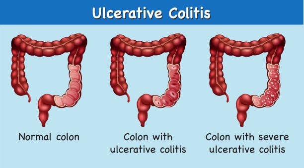
Inflammatory Bowel Disease (IBD) encompasses a group of chronic inflammatory conditions affecting the gastrointestinal tract. The two primary forms, Ulcerative Colitis (UC) and Crohn’s Disease (CD), particularly when affecting the colon, share some overlapping symptoms but possess distinct characteristics in terms of history, pathology, imaging, treatment strategies, and long-term risks. Understanding these differences is crucial for accurate diagnosis, effective management, and predicting prognosis.
Overview of Inflammatory Bowel Disease (IBD)
Inflammatory Bowel Disease is characterized by chronic inflammation of the digestive tract. While the exact cause remains unclear, it is believed to involve a complex interplay of genetic predisposition, environmental factors, immune system dysfunction, and changes in the gut microbiome. UC and CD are the most common forms, impacting millions worldwide. Although they both cause inflammation and can lead to severe complications, their patterns of disease distribution and tissue involvement differ significantly, especially when considering colonic disease.
Differentiating Features of Ulcerative Colitis and Crohn’s Disease
Distinguishing between UC and CD is fundamental in IBD management. The differentiation is typically based on a combination of clinical presentation, endoscopic findings, histopathology, and imaging studies.
Differentiating Feature 1: History and Clinical Presentation
While symptoms can overlap, certain patterns are more suggestive of one condition over the other.
-
- Ulcerative Colitis: Typically presents with bloody diarrhea, often containing mucus and pus. Other symptoms include urgency to defecate, tenesmus (feeling of incomplete evacuation), abdominal pain (often crampy and relieved by defecation), weight loss, fatigue, and fever during flares. The onset is usually gradual, but can be abrupt. UC symptoms are often continuous once established, though severity fluctuates.
- Crohn’s Disease of the Colon: Can present with non-bloody diarrhea, abdominal pain (often more generalized or right lower quadrant pain), weight loss, low-grade fever, and fatigue. While bloody diarrhea can occur if the rectum is involved, it is less common and typically less prominent than in UC. CD is also associated with extraintestinal manifestations (like joint pain, skin problems, eye inflammation) and complications like strictures, fistulas, and abscesses, which may be presenting symptoms. The clinical course is often relapsing and remitting.
Differentiating Feature 2: Pathology (Macroscopic and Microscopic)
Pathology provides some of the most definitive distinctions between UC and CD.
-
- Ulcerative Colitis: Characterized by continuous inflammation that begins in the rectum and extends proximally through the colon. It is typically limited to the mucosa and submucosa layers of the colon wall.
- Macroscopic: The colon mucosa appears erythematous, friable, and ulcerated, often with pseudopolyps (islands of regenerating mucosa). The inflammation is uniform and uninterrupted in affected areas. The rectum is almost always involved.
- Microscopic: Histology shows diffuse inflammation in the mucosa and submucosa, crypt abscesses (neutrophils accumulating in crypts), crypt distortion and branching, goblet cell depletion, and inflammatory infiltrates (lymphocytes, plasma cells, neutrophils). Granulomas (collections of macrophages) are absent in UC.
- Crohn’s Disease of the Colon: Characterized by discontinuous (skip lesions) inflammation that can affect any part of the gastrointestinal tract from mouth to anus, including the colon. Inflammation is transmural, affecting all layers of the bowel wall (mucosa, submucosa, muscularis propria, serosa).
- Macroscopic: Inflammation is patchy, with normal-appearing mucosa interspersed between affected areas. Ulcers are often deep, linear, and fissure-like, contributing to the characteristic “cobblestoned” appearance of the mucosa. Thickening of the bowel wall is common due to edema, inflammation, and fibrosis. Strictures (narrowing) and fistulas (abnormal connections between bowel segments, other organs, or skin) are hallmarks.
- Microscopic: Histology shows transmural inflammation with lymphoid aggregates. Crypt architecture distortion can be present. Non-caseating granulomas are classic but not always found (present in about 30-60% of cases) and, when present, are highly suggestive of CD. Fissuring ulcers extending deeply into the wall are also characteristic.
- Indeterminate Colitis: In about 10-15% of cases, the pathological features are not definitively UC or CD, leading to a diagnosis of indeterminate colitis. With time and further evaluation, most cases are later classified as either UC or CD.
- Ulcerative Colitis: Characterized by continuous inflammation that begins in the rectum and extends proximally through the colon. It is typically limited to the mucosa and submucosa layers of the colon wall.
Differentiating Feature 3: Imaging (X-ray Findings)
Radiological studies, particularly barium contrasts (barium enema) and cross-sectional imaging (CT or MRI), can reveal distinctive patterns.
-
- Ulcerative Colitis (Barium Enema): Shows diffuse, fine, and continuous granularity of the mucosa early on. In chronic disease, loss of haustrations (the normal sacculations of the colon) leads to a characteristic “lead pipe” or “hose-pipe” appearance of the colon, indicating a rigid, shortened tube. Ulcerations may be visible. The terminal ileum is typically normal (though mild backwash ileitis can occur).
- Crohn’s Disease of the Colon (Barium Studies/CT/MRI): Shows discontinuous or “skip” lesions. Cobblestoning of the mucosa, deep ulcers, strictures (often long and irregular), thickening of the bowel wall, and evidence of penetrating disease such as fistulas (e.g., entero-enteric, entero-colic, perianal) or abscesses are characteristic findings on barium studies and cross-sectional imaging. Fat wrapping (creeping fat around the intestine) is also seen on CT/MRI.
Differentiating Feature 4: Treatment (Medical Management)
While many drug classes are used for both conditions, the emphasis and efficacy of specific agents can differ.
- Ulcerative Colitis: Aminosalicylates (5-ASA drugs like sulfasalazine, mesalamine) are often the first-line treatment for mild-to-moderate UC and for maintaining remission, particularly in distal disease. Corticosteroids are used to induce remission in moderate-to-severe flares. Immunomodulators (like azathioprine, 6-mercaptopurine) and biologic agents (anti-TNF like infliximab, adalimumab; anti-integrin like vedolizumab; anti-IL like ustekinumab) are used for moderate-to-severe or refractory disease and for maintenance. Janus kinase (JAK) inhibitors (like tofacitinib, upadacitinib) represent newer options, particularly for moderate-to-severe UC.
- Crohn’s Disease of the Colon: 5-ASA drugs are generally less effective than in UC, though they may have some role in mild colonic CD. Corticosteroids are used for inducing remission. Immunomodulators play a significant role in maintaining remission and reducing the need for steroids, particularly for moderate-to-severe disease. Biologic agents (same classes as in UC, but selection may vary based on presentation, e.g., anti-TNF for perianal disease) are crucial for moderate-to-severe or complicated CD. Methotrexate is an option for CD (less so for UC). Nutritional therapy (e.g., exclusive enteral nutrition in pediatric CD) can also be an important adjunctive treatment.
In essence, medical therapy aims to induce and maintain remission, manage symptoms, and prevent complications in both conditions, but the specific drug choice and sequence may be guided by the disease classification, severity, and location.
Differentiating Feature 5: Risk of Cancer
Both UC and CD involving the colon increase the risk of colorectal cancer (CRC) compared to the general population, particularly with long-standing disease.
-
- Ulcerative Colitis: The risk of CRC in UC is well-established and correlates with the duration, extent, and severity of colitis. Patients with pancolitis (involvement of the entire colon) have the highest risk. Surveillance colonoscopy with multiple biopsies to detect dysplasia is standard practice, typically starting 8-10 years after diagnosis for extensive colitis.
- Crohn’s Disease of the Colon: The risk of CRC is also increased in CD involving the colon, though possibly slightly lower than in extensive UC. The risk is associated with the extent and duration of colonic involvement, presence of strictures, and potentially fistulas. Surveillance colonoscopy is also recommended for patients with colonic CD, following similar guidelines to UC. CRC in CD is often found in diseased segments, including strictures, making detection more challenging.
Role of Surgery in Managing UC and CD Complications
Surgery is a critical treatment modality in IBD, typically reserved for managing complications, disease refractory to medical therapy, or dysplasia/cancer. The role and goals of surgery differ significantly between UC and CD.
- Surgery in Ulcerative Colitis: Surgery is potentially curative for the colonic disease in UC because the disease is limited to the colon. Indications include refractory disease despite optimal medical therapy, toxic megacolon (acute dilatation of the colon with systemic toxicity), perforation, severe and uncontrollable bleeding, and high-grade dysplasia or confirmed CRC. The most common definitive surgical procedure is a total colectomy with ileal pouch-anal anastomosis (IPAA), which removes the colon and rectum while preserving bowel continuity and anal function. In some cases, a total colectomy with end ileostomy or ileorectal anastomosis may be performed.
- Surgery in Crohn’s Disease: Surgery is not curative for CD, as the disease can recur in other parts of the gastrointestinal tract or adjacent to surgical joins (anastomoses). Surgery aims to manage complications and improve quality of life. Indications include refractory strictures causing obstruction, medically resistant fistulas and abscesses, perforation, severe bleeding, and dysplasia or CRC within a diseased segment. Surgical procedures are typically segmental resections (removing only the affected segment), strictureplasty (widening a stricture without removing the bowel segment, used for multiple strictures), or drainage of abscesses. While surgery can provide significant relief from complications, re-operation rates are high due to disease recurrence.
Nonoperative Therapy of UC and CD
Nonoperative therapy forms the cornerstone of IBD management, focusing on controlling inflammation, relieving symptoms, preventing complications, and maintaining remission. This involves a combination of pharmacologic agents, nutritional support, and lifestyle modifications.
- Pharmacologic Therapy: Various drug classes are used, including aminosalicylates, corticosteroids, immunomodulators, biologic agents, and small molecule inhibitors. The choice of medication depends on disease activity, location, severity, previous treatments, and individual patient factors. Treatment strategies often involve an induction phase to achieve remission and a maintenance phase to prevent relapse.
- Nutritional Support: Nutritional deficiencies are common in IBD due to poor intake, malabsorption, increased losses, and inflammation. Nutritional assessment and support are vital. This can include dietary modifications (e.g., low-residue diets during flares, avoiding triggering foods), treating specific deficiencies (iron, B12, vitamin D), and sometimes employing enteral or parenteral nutrition, especially in malnourished CD patients or children. Exclusive enteral nutrition is a primary induction therapy in pediatric CD.
- Symptomatic Treatment: Medications to control diarrhea (e.g., loperamide), manage pain (though NSAIDs should generally be avoided as they can worsen inflammation), or treat associated conditions like anemia or bone loss are often part of the overall management plan.
- Lifestyle Modifications: Stress management techniques, regular exercise (when feasible), smoking cessation (smoking significantly worsens CD and increases its risk), and maintaining a healthy diet can contribute to better disease control and overall well-being.
Conclusion
Ulcerative Colitis and Crohn’s Disease of the colon, while both forms of IBD, exhibit critical differences in their patterns of inflammation, depth of tissue involvement, presence of granulomas, macroscopic and radiological appearances, and response to specific therapies. Accurately differentiating these conditions through clinical history, endoscopy, biopsy, and imaging is paramount for guiding management. Nonoperative medical therapy is the primary approach for both, aiming to control inflammation and maintain remission, utilizing a range of pharmacologic agents selected based on the specific diagnosis and disease characteristics. Surgery plays a vital, albeit different, role: it is potentially curative for the colonic component of UC but is typically reserved for managing complications and is not curative in CD. A multidisciplinary approach involving gastroenterologists, surgeons, radiologists, pathologists, dietitians, and mental health professionals is essential for providing comprehensive care to patients with these complex chronic conditions. Ongoing surveillance for colorectal cancer is crucial for patients with long-standing colonic involvement in both UC and CD.


