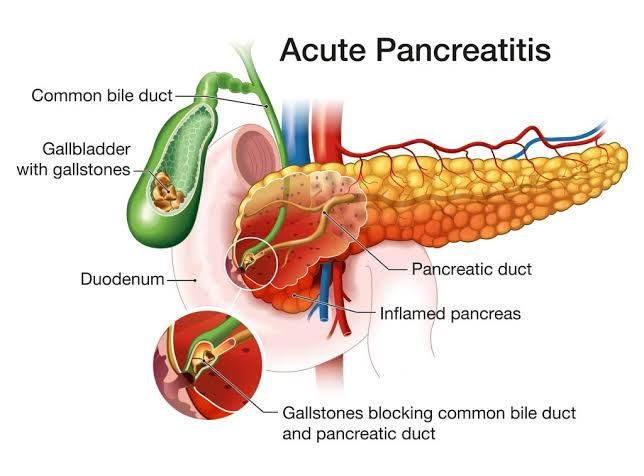
The pancreas is a vital organ playing a dual role in digestion (exocrine function, producing enzymes) and metabolism (endocrine function, producing hormones like insulin). Given its complex development and critical physiological roles, the pancreas can be affected by a range of conditions, from congenital abnormalities present at birth to acquired inflammatory diseases and aggressive malignancies. Understanding these diverse pathologies is crucial for diagnosis, treatment, and research.
Main Congenital Anomalies of the Pancreas
Congenital anomalies of the pancreas result from errors during embryonic development, specifically the fusion of the dorsal and ventral pancreatic buds. While many are asymptomatic, some can lead to significant clinical issues due to obstruction or abnormal function.
The main congenital anomalies include:
- Annular Pancreas: This is the most clinically significant anomaly. It occurs when the ventral pancreatic bud fails to rotate properly and fuses with the dorsal bud in a way that forms a ring of pancreatic tissue completely or partially encircling the second part of the duodenum.
- Clinical Significance: The constricting ring of pancreatic tissue can cause partial or complete duodenal obstruction, leading to symptoms like vomiting (especially in infants), feeding difficulties, and abdominal distension. It is sometimes associated with other congenital anomalies like Down syndrome.
- Pancreas Divisum: This is the most common congenital anomaly, present in up to 10% of the population. It results from the failure of the dorsal and ventral pancreatic ducts to fuse during development. Consequently, the bulk of the pancreatic secretion (from the dorsal pancreas via the duct of Santorini) drains through the minor duodenal papilla, which is typically smaller than the major papilla used by the ventral pancreas (via the duct of Wirsung).
- Clinical Significance: While often asymptomatic, pancreas divisum is considered a risk factor for recurrent acute pancreatitis in some individuals. The minor papilla may be too small to handle the large volume of dorsal pancreatic secretions, leading to outflow obstruction and inflammation.
- Ectopic (Accessory) Pancreas: This refers to the presence of pancreatic tissue outside the normal confines of the pancreas, without vascular or anatomical connection to the main gland. Common locations include the wall of the stomach, duodenum, jejunum, Meckel’s diverticulum, ileum, or esophagus.
- Clinical Significance: Most ectopic pancreatic rests are asymptomatic. However, they can cause symptoms depending on their location (e.g., bleeding, obstruction, or pain from inflammation) or complications like peptic ulceration in adjacent mucosa. They can also undergo pathological processes similar to the main pancreas, including inflammation or neoplastic change.
- Pancreatic Agenesis/Hypoplasia: This is a rare anomaly involving the complete absence (agenesis) or underdevelopment (hypoplasia) of the pancreas, either partially or totally. Complete agenesis is incompatible with life unless managed aggressively. Partial agenesis, often affecting the dorsal pancreas, can occur.
- Clinical Significance: Total agenesis results in severe diabetes mellitus (lack of insulin) and malabsorption (lack of exocrine enzymes) from birth. Partial agenesis can lead to varying degrees of endocrine and exocrine insufficiency.
- Congenital Pancreatic Cysts: These are rare, true cysts (lined by epithelium) thought to arise from abnormal development of pancreatic ducts. They are usually solitary, small, and located anywhere in the gland.
- Clinical Significance: Most are asymptomatic and discovered incidentally. Large cysts can cause symptoms due to mass effect. It is crucial to differentiate congenital cysts from pseudocysts or neoplastic cysts.
Cystic Fibrosis and its Etiology, Pathogenesis, and Pathologic Features
Cystic Fibrosis (CF) is an autosomal recessive genetic disorder that affects multiple organs, most notably the lungs, pancreas, gastrointestinal tract, sweat glands, and genitourinary tract. Pancreatic involvement is one of the most common manifestations.
- Definition: Cystic fibrosis is a monogenic disorder caused by mutations in the gene encoding the Cystic Fibrosis Transmembrane Conductance Regulator (CFTR) protein. CFTR is an epithelial chloride ion channel and also regulates other ion channels. Its dysfunction leads to abnormal transport of chloride and sodium ions across epithelial membranes, resulting in dehydrated, thick, and viscous secretions in various exocrine glands.
- Etiology: The cause of CF is a mutation in the CFTR gene located on chromosome 7q31.2. Over 2,000 different mutations have been identified, but the most common is a deletion of three base pairs resulting in the absence of phenylalanine at position 508 (ΔF508). CF is inherited in an autosomal recessive pattern, meaning an individual must inherit two mutated copies of the CFTR gene (one from each parent) to have the disease. Individuals with one mutated copy are carriers and typically asymptomatic.
- Pathogenesis: The defective or absent CFTR protein disrupts ion and water transport in epithelial cells, particularly those lining ducts of exocrine glands. In the pancreas, CFTR is crucial for regulating bicarbonate and chloride secretion into the pancreatic ducts. Without functional CFTR, bicarbonate secretion is impaired, leading to acidic and concentrated pancreatic fluid. This acidic, low-volume fluid causes precipitation of proteins within the ducts. The thick, viscous secretions obstruct the small pancreatic ducts.This ductal obstruction leads to:
- Stasis of pancreatic enzymes.
- Swelling of the acinar cells secondary to blocked outflow.
- Premature activation of pancreatic enzymes within the acinar cells due to back pressure and cellular damage.
- Autodigestion of pancreatic tissue.
- Chronic inflammation and fibrosis as the body attempts to repair the damage.
The severity of pancreatic involvement correlates with the degree of CFTR dysfunction, which in turn depends on the specific CFTR mutations. Some mutations lead to almost complete loss of function and severe pancreatic insufficiency, while others result in partial function and pancreatic sufficiency (where the pancreas retains some exocrine function).
- Pathologic Features:
- Macroscopic: In severe, long-standing cases (typical in pancreatic insufficiency), the pancreas is significantly smaller and firmer than normal. It may appear fatty and fibrotic. Cysts and ductal dilation may be visible.
- Microscopic: The hallmark feature is severe atrophy and progressive loss of exocrine acinar tissue, which is replaced by dense fibrous tissue and fat. The pancreatic ducts are often dilated and filled with eosinophilic, inspissated (thickened) protein plugs or calcifications. Chronic inflammatory cells (lymphocytes, plasma cells) are present in the fibrous stroma. The islets of Langerhans (endocrine tissue) are often relatively preserved initially but can eventually be affected, leading to CF-related diabetes. In individuals with pancreatic sufficiency, the changes are milder, potentially involving only focal ductal dilation and mild fibrosis.
The Causes, Pathogenesis, and Pathologic Features of Different Forms of Pancreatitis
Pancreatitis is inflammation of the pancreas. It occurs when digestive enzymes produced by the pancreas become activated prematurely within the gland itself, leading to autodigestion and inflammation. Pancreatitis can be acute (sudden onset, often resolving) or chronic (persistent inflammation and irreversible damage).
Acute Pancreatitis:
- Definition: An acute inflammatory process of the pancreas with variable involvement of other regional tissues or distant organ systems.
- Causes: The two most common causes account for about 80% of cases:
- Gallstones: Small gallstones obstructing the common bile duct and/or main pancreatic duct at the ampulla of Vater, causing reflux of bile or pancreatic juice into the pancreatic duct and increased intraductal pressure.
- Alcohol Abuse: Chronic heavy alcohol consumption is a major risk factor and cause. The exact mechanism is complex but likely involves potentiation of zymogen activation, direct toxic effects on acinar cells, oxidative stress, and possibly increased protein plug formation in ducts. Other causes include:
- Hypertriglyceridemia (very high triglyceride levels)
- Certain medications (e.g., thiazide diuretics, azathioprine)
- Post-Endoscopic Retrograde Cholangiopancreatography (ERCP)
- Trauma (e.g., blunt abdominal injury)
- Viral infections (e.g., mumps, coxsackie)
- Hypercalcemia
- Genetic factors (e.g., hereditary pancreatitis caused by mutations in genes like PRSS1, SPINK1)
- Idiopathic (no identifiable cause, about 10-15% of cases)
- Pathogenesis: The central event is the inappropriate activation of pancreatic enzymes within the acinar cells rather than in the intestinal lumen.
- Triggering events: Acinar cell injury (e.g., from toxins like alcohol, ischemia), ductal obstruction (e.g., gallstones, plugs), or defective intracellular transport of zymogens (inactive enzyme precursors) can initiate the process.
- Enzyme activation: Key enzymes like trypsinogen are prematurely converted to active trypsin. Trypsin then activates other zymogens (proelastase, procarboxypeptidase) and prophospholipase A2.
- Autodigestion: Activated enzymes attack pancreatic tissue:
- Trypsin digests proteins.
- Elastase degrades elastic fibers in blood vessels, leading to hemorrhage.
- Phospholipase A2 degrades phospholipids in cell membranes, causing cell necrosis.
- Lipase hydrolyzes triglycerides in fat cells, causing fat necrosis.
- Inflammation: Tissue damage triggers an intense inflammatory response, attracting leukocytes and releasing pro-inflammatory cytokines (e.g., TNF-α, IL-1, IL-6). This can lead to systemic inflammatory response syndrome (SIRS) and organ failure.
- Pathologic Features:
- Macroscopic: The appearance varies with severity. In mild interstitial edematous pancreatitis, the gland is enlarged and edematous. In severe necrotizing pancreatitis, there are areas of chalky-white fat necrosis (due to saponification of fat by lipase), hemorrhagic areas (due to vascular damage by elastase and trypsin), and gray-white foci of enzymatic parenchymal necrosis.
- Microscopic: Key features include interstitial edema, inflammatory infiltrate (neutrophils in early stages, later mixed inflammatory cells), fat necrosis (shadows of necrotic adipocytes surrounded by inflammatory cells), and variable degrees of parenchymal necrosis (acinar and ductal cell death). Vascular injury and thrombosis can also be seen, contributing to ischemic necrosis.
Chronic Pancreatitis:
- Definition: A prolonged process of inflammation of the pancreas characterized by irreversible destruction of the exocrine parenchyma, fibrosis, and eventually, in later stages, destruction of the endocrine parenchyma.
- Causes:
- Chronic Alcohol Abuse: This is the most common cause globally. Chronic alcohol consumption is thought to cause repeated episodes of subclinical or clinical acute pancreatitis, leading to progressive damage. Mechanisms may involve toxic effects, oxidative stress, and protein plug formation in ducts.
- Idiopathic (no identifiable cause): A significant proportion, sometimes referred to as tropical pancreatitis in certain geographic areas.
- Severe or Recurrent Acute Pancreatitis: Damage from previous acute episodes.
- Hereditary Pancreatitis: Mutations in genes like PRSS1 (cationic trypsinogen), SPINK1 (serine protease inhibitor), or CFTR.
- Autoimmune Pancreatitis: A distinct form characterized by autoimmune inflammation and often responding to steroids.
- Obstructive Chronic Pancreatitis: Due to prolonged obstruction of the main pancreatic duct (e.g., by stones, strictures, or tumors).
- Pathogenesis: Unlike acute pancreatitis, the pathogenesis is complex and often involves repeated cycles of injury, inflammation, and repair leading to progressive fibrosis. The precise mechanisms are debated but likely involve:
- Acinar cell injury initiating inflammatory cascades.
- Activation of pancreatic stellate cells, which are the primary source of extracellular matrix proteins leading to fibrosis.
- Ductal obstruction by protein plugs (especially in alcoholic pancreatitis) or strictures contributing to upstream damage.
- Inflammatory mediators perpetuating the fibrotic response.
- Oxidative stress.
- Pathologic Features:
- Macroscopic: The gland is typically smaller, harder, and irregular due to extensive fibrosis. The pancreatic ducts are often dilated, strictured, and may contain calcifications (pancreaticolithiasis), which are common, especially in alcoholic chronic pancreatitis. Pseudocysts (collections of fluid surrounded by inflammatory tissue, not true cysts) may be present.
- Microscopic: Characteristic features include widespread loss of acinar tissue replaced by dense fibrous tissue. There is chronic inflammation with lymphocytes, plasma cells, and macrophages. The pancreatic ducts are dilated, irregular, and lined by flattened or hyperplastic epithelium; protein plugs or calcifications may be seen within the lumens. Islets of Langerhans appear relatively spared in early stages dispersed within the fibrosis, but eventually, they can become entrapped and damaged, leading to impaired insulin and glucagon production and diabetes mellitus.
Major Tumors of the Exocrine Pancreas
Tumors can arise from both the exocrine and endocrine components of the pancreas. Tumors of the exocrine pancreas are far more common and generally have a worse prognosis than endocrine tumors. The vast majority originate from ductal epithelial cells.
The major tumor of the exocrine pancreas is:
- Pancreatic Ductal Adenocarcinoma (PDAC): This is the most common type of pancreatic cancer, accounting for over 85% of malignant pancreatic neoplasms. It is a highly aggressive malignancy with a poor prognosis, largely due to late diagnosis and early metastasis.
- Description: PDAC is an adenocarcinoma arising from the epithelial cells lining the pancreatic ducts. These tumors typically induce a marked desmoplastic stromal reaction (growth of fibrous tissue), which contributes to their hardness and imaging characteristics.
- Location: Most PDACs occur in the head of the pancreas (about 60-70%), followed by the body (15-20%) and tail (10-15%). Tumors in the head often cause symptoms earlier by obstructing the common bile duct (leading to jaundice) or pancreatic duct.
- Risk Factors: Major risk factors include smoking (doubles the risk), chronic pancreatitis (especially hereditary or long-standing alcoholic), older age, diabetes mellitus (both long-standing and new-onset can be a symptom), obesity, certain germline genetic mutations (e.g., BRCA2, PALB2, ATM, Lynch syndrome genes, Peutz-Jeghers syndrome – STK11), and exposure to certain chemicals.
- Pathogenesis: PDAC is believed to arise from precursor lesions, primarily Pancreatic Intraepithelial Neoplasia (PanIN). PanINs are microscopic, flat or papillary epithelial proliferations within small ducts, graded from low-grade (PanIN-1) to high-grade (PanIN-3, carcinoma in situ). The progression from normal epithelium through low-grade to high-grade PanIN and ultimately invasive adenocarcinoma is a multi-step process driven by accumulating genetic mutations (common mutations include KRAS, TP53, CDKN2A/p16, and SMAD4/DPC4). Other precursor lesions include Intraductal Papillary Mucinous Neoplasms (IPMNs) and Mucinous Cystic Neoplasms (MCNs), which are larger, macroscopic cystic lesions with malignant potential.
- Pathologic Features:
- Macroscopic: Typically presents as a firm, ill-defined, gray-white mass. In the head, it often infiltrates surrounding tissues, including the bile duct, duodenum, and major vessels.
- Microscopic: Characterized by infiltrating glands and nests of malignant epithelial cells embedded within a dense, abundant desmoplastic stroma (fibrous tissue). The glands are often irregular in shape and size, with crowded, atypical nuclei, and scant cytoplasm. Perineural invasion (tumor cells infiltrating nerves) and vascular invasion are common microscopic features contributing to the aggressive nature and propensity for local spread and metastasis. Tumor cells may produce mucin.
While PDAC is the major exocrine malignancy, it’s worth noting other less common neoplastic lesions derived from exocrine epithelium, such as:
- Intraductal Papillary Mucinous Neoplasms (IPMNs): Mucus-producing epithelial neoplasms arising from pancreatic ducts. They are classified by the type of duct involved (main duct, branch duct, or mixed) and by grade of dysplasia (low, intermediate, high, invasive carcinoma). Main-duct IPMNs have a higher risk of progression to invasive carcinoma than typical branch-duct IPMNs.
- Mucinous Cystic Neoplasms (MCNs): Cystic neoplasms typically occurring in the body or tail of the pancreas, almost exclusively in women. They consist of mucin-producing cysts lined by columnar epithelium and surrounded by a characteristic ovarian-type stroma. Like IPMNs, they have malignant potential depending on the degree of epithelial dysplasia.
- Solid Pseudopapillary Neoplasm (SPPN): A rare, low-grade malignant tumor that typically affects young women. It has solid and pseudopapillary areas microscopically.
These cystic neoplasms are important because they can harbor or develop into invasive adenocarcinoma, but PDAC remains the defining “major tumor” due to its prevalence and lethality.


