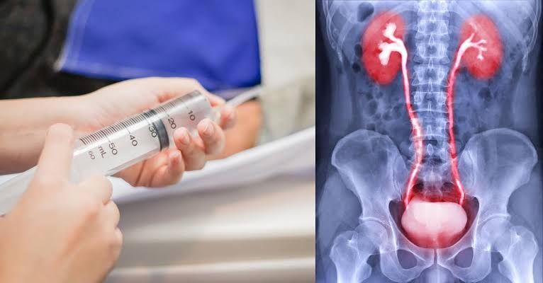
Diagnostic imaging plays a critical role in modern medicine, enabling healthcare professionals to visualize internal structures and identify abnormalities. While techniques like X-ray, Computed Tomography (CT), Magnetic Resonance Imaging (MRI), and Ultrasound provide valuable information, their diagnostic yield can often be significantly enhanced through the use of contrast media.
Contrast media, also referred to as contrast agents, are pharmacological substances introduced into the body to alter the way tissues or structures appear on diagnostic images. They work by transiently changing the signal intensity or attenuation of specific areas, making blood vessels, organs, or lesions more visible against surrounding tissues.
Types of Contrast Media (Pharmacological Agents for Diagnostic Purposes)
Contrast media are broadly classified based on the imaging modality they are designed to enhance and their chemical composition. The primary types used in clinical practice include iodinated contrast agents, gadolinium-based contrast agents, and ultrasound contrast agents.
1. Iodinated Contrast Media (for X-ray and CT)
Iodinated contrast media are the most commonly used type, particularly in X-ray and CT imaging. They contain iodine atoms, which effectively absorb X-rays, leading to increased attenuation in the areas where they accumulate (e.g., blood vessels, kidneys, bladder). This results in a bright signal on the resulting image.
- Mechanism: When injected intravenously, iodinated contrast media distribute throughout the bloodstream, highlighting blood vessels (angiography) and perfused tissues. They are primarily eliminated via glomerular filtration in the kidneys, allowing visualization of the renal system (urography or nephrography). Oral or rectal administration is used to opacify the gastrointestinal tract.
- Classification (Historically & Modern Use):
- Ionic vs. Non-ionic: Early contrast media were ionic, dissociating into charged particles in solution. Non-ionic agents remain non-dissociated molecules. Non-ionic agents are generally preferred in modern practice due to a lower incidence of adverse reactions, likely related to lower osmolality and chemical toxicity.
- High-Osmolar vs. Low-Osmolar vs. Iso-Osmolar: Osmolality refers to the concentration of solute particles in a solution. High-osmolar contrast media (HOCM) have an osmolality significantly higher than blood plasma. Low-osmolar contrast media (LOCM) have an osmolality closer to plasma, and iso-osmolar contrast media (IOCM) have an osmolality equal to plasma. LOCM and IOCM are associated with fewer uncomfortable side effects (like heat sensation or pain on injection) and a lower risk of certain adverse reactions compared to HOCM, leading to their widespread adoption.
- Administration Routes: Intravenous (most common), Oral, Rectal, Intra-articular, Intrathecal, etc.
2. Gadolinium-Based Contrast Agents (GBCAs) (for MRI)
GBCAs are paramagnetic substances used as contrast agents in Magnetic Resonance Imaging (MRI). Gadolinium itself is toxic as a free ion, so it is chelated (bound) to an organic molecule to form a stable complex that can be safely administered.
- Mechanism: GBCAs alter the local magnetic field created by the MRI scanner, primarily affecting the T1 relaxation time of surrounding water protons. This leads to increased signal intensity (brightness) in areas where the agent accumulates or perfuses, making lesions, inflammation, or vascular structures more conspicuous on T1-weighted MRI images.
- Classification (by Structure):
- Linear: Characterized by an open, linear molecular structure where the gadolinium ion is chelated. Some linear agents have been associated with a higher risk of Nephrogenic Systemic Fibrosis (NSF) in patients with severe renal impairment.
- Macrocyclic: Feature a cage-like structure tightly enclosing the gadolinium ion. Macrocyclic agents are considered more stable than linear agents and are associated with a lower risk of dissociation and subsequent toxicity, including NSF. Regulatory bodies often prefer or recommend the use of macrocyclic over linear agents, especially in higher-risk patients.
- Administration Route: Primarily Intravenous.
- Elimination: Primarily via renal excretion.
3. Ultrasound Contrast Agents (UCAs) (for Ultrasound)
UCAs, often referred to as microbubble contrast agents, are gas-filled microbubbles surrounded by a stabilizing shell (e.g., lipid, polymer, protein).
- Mechanism: When injected intravenously, these microbubbles circulate within the bloodstream. They oscillate and resonate when exposed to ultrasound waves, generating strong echoes that are detected by the ultrasound transducer. This enhances the visualization of blood flow (perfusion) in organs and allows for better delineation of lesions. The microbubbles are small enough to pass through pulmonary capillaries but are too large to pass through the intact endothelium of capillaries in most tissues, thus remaining purely intravascular.
- Composition: Typically consist of high molecular weight gases (e.g., sulfur hexafluoride, perflutren) which are poorly soluble in blood, contained within a flexible shell.
- Administration Route: Intravenous.
- Elimination: The gas is exhaled, and the shell components are metabolized.
4. Other Contrast Agents
While the above are the most common injectable agents, other contrast media are used for specific purposes:
- Barium Sulfate (for X-ray/CT of GI Tract): An insoluble salt that is orally or rectally administered to coat and opacify the lumen of the gastrointestinal tract. It strongly absorbs X-rays.
- Oral Contrast for CT (Dilute Iodinated Solutions): Dilute solutions of iodinated contrast are sometimes used orally to opacify the GI tract for CT scans, especially in patients where barium is contraindicated or aspiration is a concern.
Adverse Reactions of Contrast Media
While generally safe, the administration of contrast media carries a risk of adverse reactions, ranging from minor to severe. It is crucial for healthcare professionals to anticipate, recognize, and manage these reactions promptly.
Adverse reactions can be broadly categorized by their timing and mechanism:
- Acute Reactions: Occur rapidly, typically within minutes to an hour after administration.
- Minor: Nausea, vomiting, warmth sensation, flushing, mild urticaria (hives), itching, headache, dizziness. These are usually self-limiting and require minimal or no treatment beyond observation and reassurance.
- Moderate: More widespread urticaria, moderate bronchospasm, mild facial or laryngeal edema, moderate hypotension, vagal reactions (bradycardia, hypotension, syncope). Require active management and closer monitoring.
- Severe (Anaphylactoid/Anaphylactic): Life-threatening reactions resembling anaphylaxis. Severe bronchospasm, significant laryngeal edema leading to airway compromise, profound hypotension, shock, arrhythmias, cardiovascular collapse, cardiac arrest. These require immediate, aggressive intervention.
- Delayed Reactions: Occur hours to days after administration.
- Most commonly include skin reactions (e.g., maculopapular rash). Flu-like symptoms, fatigue, or other mild systemic effects can also occur. These are usually self-limiting but may require symptomatic treatment.
- Chemically Mediated Reactions: Related to the inherent properties of the contrast agent (e.g., osmolality, volume, calcium binding). Examples include warmth, pain on injection (more common with HOCM), nausea, and vomiting.
- Organ-Specific Toxicity:
- Contrast-Induced Nephropathy (CIN): Acute kidney injury following contrast administration (discussed in Section 4).
- Nephrogenic Systemic Fibrosis (NSF): A rare but serious fibrosing disorder affecting the skin and internal organs, linked to certain linear GBCAs in patients with severe renal dysfunction.
- Thyroid Dysfunction: Iodinated contrast can affect thyroid function, particularly in patients with pre-existing thyroid disease or neonates/infants.
Risk Factors for Adverse Reactions:
- History of previous reaction to contrast media (especially moderate or severe).
- History of other allergies (asthma, hay fever, food allergies – though this link is debated, it warrants consideration).
- Anxiety.
- Beta-blocker medication use (can worsen bronchospasm and blunt response to epinephrine).
- Interleukin-2 therapy.
- Certain medical conditions (e.g., asthma, severe cardiac disease, pheochromocytoma).
Management of Adverse Reactions
Effective management of contrast media reactions relies on preparedness, prompt recognition, and appropriate intervention guided by the severity of the reaction.
Key Principles of Management:
- Preparedness:
- Maintain a well-stocked emergency cart with essential medications (epinephrine, antihistamines, corticosteroids, bronchodilators, IV fluids) and equipment (oxygen, airway supplies, monitoring devices).
- Ensure all staff involved in contrast administration are trained in recognizing and managing acute reactions, including basic and, ideally, advanced cardiac life support (BLS/ACLS).
- Have established protocols for managing reactions readily available.
- Recognition and Assessment:
- Continuously monitor the patient during and immediately after contrast injection.
- Rapidly assess the severity of any reaction (minor, moderate, severe).
- Look for objective signs (e.g., rash extent, presence of wheezing, blood pressure changes, airway swelling).
- Intervention (Based on Severity):
- Minor Reactions (e.g., mild urticaria, nausea):
- Stop contrast infusion.
- Observe and reassure the patient.
- Administer oral or IV antihistamines (e.g., diphenhydramine) if urticaria/itching is bothersome.
- Monitor vital signs and symptoms.
- Moderate Reactions (e.g., widespread urticaria, mild bronchospasm, mild hypotension):
- Stop contrast infusion.
- Elevate legs if hypotensive.
- Administer oxygen if needed.
- Administer IV antihistamines (e.g., diphenhydramine).
- Administer IV corticosteroids (e.g., methylprednisolone) – beneficial for preventing delayed reactions, but onset of action is slow for acute symptoms.
- Administer inhaled bronchodilators (e.g., albuterol) for bronchospasm.
- Monitor vital signs closely. May require IV fluids for hypotension.
- Severe Reactions (Anaphylactoid/Anaphylactic – e.g., severe bronchospasm, laryngeal edema, profound hypotension): This is an emergency.
- Call for immediate assistance/Code Team.
- Lay the patient flat, elevate legs.
- Administer oxygen at a high flow rate.
- ** administer Epinephrine (Adrenaline):** This is the single most important treatment.
- Intramuscular (IM) Epi: Preferred initial route for systemic reactions with hypotension/airway compromise/diffuse urticaria. Administer 0.3-0.5 mg of 1:1000 epinephrine IM into the lateral thigh. Can repeat every 5-15 minutes as needed.
- Intravenous (IV) Epi: Reserved for severe, refractory hypotension or cardiovascular collapse. Requires careful titration and monitoring (e.g., 0.1 mg of 1:10,000 solution slowly IV, or continuous infusion). Requires expertise.
- Establish secure IV access; administer large volumes of IV fluids (e.g., 0.9% Saline) rapidly for hypotension.
- Administer IV antihistamines and corticosteroids.
- Manage airway if necessary (e.g., intubation for severe laryngeal edema).
- Treat bronchospasm vigorously with inhaled bronchodilators.
- Monitor ECG and vital signs continuously.
- Minor Reactions (e.g., mild urticaria, nausea):
- Documentation: Thoroughly document the timing, nature, severity of the reaction, and all interventions provided.
Methods for Reducing the Risk of Contrast-Induced Nephropathy (CIN)
Contrast-Induced Nephropathy (CIN) is an acute impairment of renal function that occurs after the administration of contrast media, typically defined as a rise in serum creatinine concentration from baseline within 48-72 hours, not attributable to other causes. It is a significant concern, especially in high-risk patients, as it can lead to prolonged hospitalization, increased morbidity, and mortality.
Reducing the risk of CIN is primarily achieved through patient assessment, optimal hydration, appropriate contrast selection and minimization, and careful management of concomitant conditions and medications.
Key Strategies for CIN Prevention:
- Identify High-Risk Patients:
- Assessment of Renal Function: This is paramount. Evaluate serum creatinine (or eGFR – estimated Glomerular Filtration Rate) in all patients undergoing contrast-enhanced studies, especially those with known risk factors. A baseline eGFR < 60 mL/min/1.73m² is a significant risk factor, with risk increasing as eGFR declines.
- Other Risk Factors: Identify patients with diabetes mellitus (especially with concurrent nephropathy), dehydration, heart failure, advanced age, patients receiving potentially nephrotoxic medications (e.g., NSAIDs, certain antibiotics), severe uncontrolled hypertension, and those undergoing large volume contrast administration or multiple contrast studies in a short timeframe.
- Ensuring Adequate Hydration:
- This is widely considered the most effective preventive measure.
- Protocolized Intravenous Hydration: Administering intravenous fluids (most commonly 0.9% Saline or Sodium Bicarbonate solution) before and after contrast administration helps dilute the contrast medium in the tubules, maintain renal blood flow, and reduce tubular injury.
- Typical Protocol (example): IV Saline 0.9% at 100-150 mL/hour starting 3-12 hours before the procedure and continuing for 6-24 hours afterward. Specific protocols vary between institutions and patient risk levels.
- Oral Hydration: Encourage adequate oral fluid intake in the 12-24 hours before the procedure for patients who can tolerate it, especially for lower-risk individuals or as an adjunct to IV hydration.
- Contrast Agent Selection and Administration:
- Use Low-Osmolar Non-ionic (LOCM) or Iso-Osmolar Non-ionic (IOCM) Contrast Media: These agents are associated with a lower risk of CIN compared to older high-osmolar ionic agents.
- Minimize Contrast Volume: Use the lowest clinically necessary dose of contrast medium. Adhere to weight-based or protocol-defined limits where appropriate.
- Minimize Repeat Exposure: Avoid repeat contrast-enhanced studies within a short period (e.g., 24-48 hours), especially in high-risk patients, unless clinically essential.
- Manage Concomitant Medications:
- Hold Nephrotoxic Medications: Temporarily discontinue potentially nephrotoxic drugs, such as NSAIDs (Non-Steroidal Anti-Inflammatory Drugs), cyclosporine, certain antibiotics (e.g., aminoglycosides), before and for a period after contrast administration in high-risk patients.
- Metformin: For patients with diabetes taking Metformin and having impaired renal function (eGFR < 30-45 mL/min), Metformin should be temporarily discontinued at the time of or prior to contrast administration and withheld for 48 hours post-procedure, or until renal function is stable, due to the risk of lactic acidosis if acute kidney injury occurs.
- ACE Inhibitors/ARBs: While some clinicians hold these medications due to theoretical concerns about reducing renal perfusion, current evidence is conflicting, and the benefit may not outweigh the risk of withholding essential blood pressure control. Institutional protocols vary.
- Other Pharmacological Interventions (Evidence Varies):
- N-acetylcysteine (NAC): While widely used historically, large randomized controlled trials and meta-analyses have yielded conflicting results, and its benefit beyond adequate hydration is uncertain, particularly with modern LOCM/IOCM agents. It is relatively inexpensive and safe, so it is sometimes used, often in combination with hydration, but it is not considered a substitute for hydration.
- Sodium Bicarbonate: Some studies suggest sodium bicarbonate hydration protocols might be superior to saline, possibly by counteracting oxidative stress, but other studies show no difference.
Conclusion
Contrast media are indispensable tools in diagnostic imaging, significantly improving the visibility of anatomical structures and pathological processes. A thorough understanding of the different types of contrast agents, their mechanisms of action, potential adverse reactions, and strategies for risk reduction is essential for all healthcare professionals involved in their administration and monitoring. By carefully identifying patient risk factors, ensuring adequate hydration, selecting appropriate agents, minimizing volumes, and being prepared to manage acute reactions, we can maximize the diagnostic benefits of contrast media while prioritizing patient safety and minimizing potential complications like contrast-induced nephropathy. Continuous education and adherence to established protocols are key to safe contrast media utilization.


