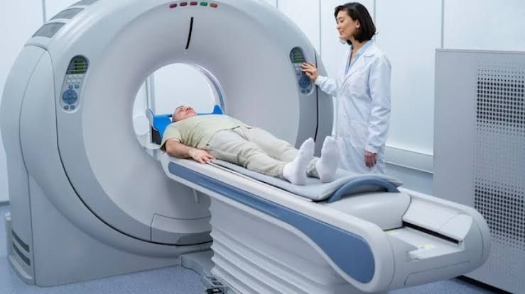
Introduction to PET Imaging
Positron Emission Tomography (PET) imaging is a non-invasive diagnostic technique that involves the use of a small amount of radioactive tracer to visualize the metabolic activity within the body. The tracer is injected into the bloodstream, where it accumulates in areas with high metabolic activity. The PET scanner detects the radiation emitted by the tracer, creating detailed images of the body’s internal structures.
How PET Imaging Works
The process of PET imaging involves several steps:
- Tracer Injection: A small amount of radioactive tracer, typically Fluorodeoxyglucose (FDG), is injected into the bloodstream. FDG is a glucose molecule that has been labeled with a radioactive isotope, usually Fluorine-18.
- Tracer Uptake: The FDG tracer is taken up by cells throughout the body, with higher uptake in areas with high metabolic activity, such as cancer cells.
- PET Scan: The patient is placed inside the PET scanner, which detects the radiation emitted by the tracer. The scanner consists of a series of detectors that surround the patient’s body.
- Image Reconstruction: The detected radiation is used to reconstruct detailed images of the body’s internal structures. The images are typically displayed in a color scale, with areas of high tracer uptake appearing as bright colors.
Understanding PET Image Interpretation
Interpreting PET images requires a thorough understanding of the underlying anatomy, physiology, and metabolic activity within the body. Here are some key factors to consider when interpreting PET images:
- Tracer Uptake: Areas with high tracer uptake indicate high metabolic activity, which can be a sign of disease, such as cancer.
- Standardized Uptake Value (SUV): SUV is a quantitative measure of tracer uptake, which helps to normalize the image data. SUV values can be used to compare the metabolic activity between different regions or patients.
- Image Fusion: PET images are often fused with other imaging modalities, such as Computed Tomography (CT) or Magnetic Resonance Imaging (MRI), to provide a more comprehensive understanding of the anatomy and metabolic activity.
Applications of PET Imaging
PET imaging has a wide range of applications in the medical field, including:
- Oncology: PET imaging is used to diagnose and monitor cancer, assess treatment response, and detect recurrence.
- Neurology: PET imaging is used to diagnose and monitor neurological disorders, such as Alzheimer’s disease, Parkinson’s disease, and epilepsy.
- Cardiology: PET imaging is used to assess myocardial viability, detect coronary artery disease, and monitor cardiac function.
Advantages and Limitations of PET Imaging
PET imaging offers several advantages over other imaging modalities, including:
- High Sensitivity: PET imaging is highly sensitive to metabolic activity, allowing for early detection of disease.
- Non-Invasive: PET imaging is a non-invasive technique, reducing the risk of complications and improving patient comfort.
However, PET imaging also has some limitations:
- Radiation Exposure: PET imaging involves exposure to ionizing radiation, which can be a concern for patients and healthcare workers.
- Cost and Availability: PET imaging is a relatively expensive and complex modality, limiting its availability in some regions.
Future Directions in PET Imaging
The field of PET imaging is continually evolving, with advances in technology and tracer development. Some future directions in PET imaging include:
- New Tracer Development: Researchers are developing new tracers that target specific diseases or biological processes, such as inflammation or immune response.
- Hybrid Imaging: Hybrid imaging modalities, such as PET/CT and PET/MRI, are becoming increasingly popular, providing a more comprehensive understanding of anatomy and metabolic activity.
- Quantitative PET Imaging: Quantitative PET imaging techniques, such as SUV and kinetic modeling, are being developed to provide more accurate and reproducible measurements of metabolic activity.
In conclusion, PET imaging is a powerful diagnostic tool that provides valuable information about metabolic activity within the body. By understanding the principles of PET imaging, interpreting PET images, and appreciating its applications and limitations, healthcare professionals can harness the full potential of this technology to improve patient care.
Fundamental Principles of PET/CT in Oncology:
-
- Metabolic Imaging: Malignant cells often exhibit increased glucose metabolism compared to normal cells, leading to higher uptake of the FDG tracer. This allows PET to identify potential tumor activity anywhere in the body.
- Whole-Body Survey: PET/CT can scan the entire body in a single session, making it highly effective for detecting distant metastases and assessing regional and distant lymph node involvement, which are crucial for accurate staging.
- Functional vs. Anatomical Information: While CT provides anatomical detail (size, shape, location), PET provides functional insight into metabolic activity. The combination (PET/CT) offers both, significantly improving diagnostic accuracy by differentiating active tumor from benign lesions, post-treatment changes (like scar tissue or necrosis), or inflammation.
- Impact on Patient Management: Accurate staging and restaging provided by PET/CT are essential for determining prognosis, selecting appropriate treatment strategies (e.g., surgery, radiation therapy, chemotherapy, targeted therapy), monitoring treatment response, and planning radiation therapy fields.
Staging and Restaging:
-
- Staging: This refers to the process of determining the extent of a cancer at the time of diagnosis. The most common system is the TNM (Tumor, Node, Metastasis) system, which describes the size and extent of the primary tumor (T), whether cancer has spread to nearby lymph nodes (N), and whether cancer has spread to distant parts of the body (M). PET/CT is particularly valuable in assessing the N (nodal) and M (metastasis) components, often revealing occult disease not visible on conventional imaging, leading to more accurate staging and appropriate treatment planning.
- Restaging: This involves reassessing the cancer status after treatment has been initiated or completed, or when recurrence is suspected. Restaging is used to:
- Evaluate response to therapy (e.g., did the tumor shrink or become less metabolically active?).
- Distinguish between active tumor and benign post-treatment changes.
- Detect residual disease after initial therapy.
- Identify sites of recurrence if the cancer returns.
- Guide treatment decisions based on the current disease status.
Applications of PET/CT in Specific Cancers (Staging and Restaging):
-
- Lung Cancer:
- Staging: PET/CT is crucial for staging non-small cell lung cancer (NSCLC), especially for assessing mediastinal and hilar lymph nodes (N staging) and detecting distant metastases (M staging). It is significantly more accurate than CT alone for identifying involved lymph nodes and occult distant disease, often preventing unnecessary surgery in patients with undetected metastases (upstaging) or allowing potentially curative treatment by accurately identifying limited disease (downstaging). Its role in small cell lung cancer (SCLC) is more focused on detecting distant disease in patients considered for limited-stage therapy.
- Restaging: Used to distinguish residual/recurrent tumor from post-treatment inflammation or scarring after surgery or chemoradiation. Also vital for monitoring response to systemic therapy and detecting recurrence in patients with rising tumor markers or clinical suspicion. Can help identify new primary lung cancers or synchronous malignancies.
- Colorectal Cancer:
- Staging: PET/CT is less sensitive for primary colon or rectal tumors, especially early-stage, or superficial regional nodes. Its main value in initial staging is in identifying distant metastatic disease (M). It is particularly useful in patients with locally advanced rectal cancer being considered for neoadjuvant therapy, or in cases where high-risk features suggest a higher likelihood of distant spread.
- Restaging: PET/CT is widely used and highly valuable for restaging colorectal cancer, particularly in patients with rising Carcinoembryonic Antigen (CEA) levels but negative or equivocal conventional imaging. It is highly effective in detecting metastases in common sites like the liver, lungs, peritoneum, bones, and extra-abdominal lymph nodes, aiding in the planning of salvage therapy or recurrence management. It helps differentiate metabolically active recurrence from scar tissue at surgical sites.
- Breast Cancer:
- Staging: The role of PET/CT in initial staging of breast cancer is generally limited to patients with locally advanced disease (Stage III) or suspected metastatic disease (Stage IV). It is not typically used for early-stage (Stage I/II) staging unless there are specific clinical concerns, as conventional imaging (mammography, ultrasound, MRI, sentinel node biopsy) is sufficient. In advanced disease, PET/CT helps detect distant metastases that may not be evident on conventional scans, impacting treatment decisions (locoregional vs. systemic therapy). Some subtypes, like invasive lobular carcinoma, can be less FDG-avid.
- Restaging: PET/CT is very effective for restaging breast cancer when recurrence or metastatic disease is suspected, or for monitoring response to systemic therapy in advanced disease. It can detect metastatic lesions in bones, liver, lungs, lymph nodes, and other sites better than some conventional methods, especially in differentiating active disease from benign conditions in the bone or after treatment.
- Oesophageal Cancer:
- Staging: PET/CT is considered standard for staging oesophageal cancer. It is highly accurate in assessing regional lymph node involvement (N) and detecting distant metastases (M) in sites like the liver, lung, bone, and distant lymph nodes. This information is critical for determining whether a patient is a candidate for curative surgery or if palliative treatment is more appropriate. PET/CT can upstage a significant number of patients compared to conventional imaging, altering management plans.
- Restaging: Plays a crucial role in restaging after neoadjuvant chemoradiation therapy, prior to planned surgery. A significant reduction in FDG uptake indicates a good metabolic response, which correlates with better pathological response and prognosis. It is also used to identify distant recurrence or progression in patients who have completed initial therapy.
- Lymphoma:
- Staging: PET/CT is the standard of care for initial staging of most FDG-avid lymphomas, including Hodgkin lymphoma and many subtypes of non-Hodgkin lymphoma (NHL), such as Diffuse Large B-cell Lymphoma (DLBCL) and Follicular Lymphoma. It is superior to CT alone for determining the full extent of nodal and extranodal involvement, accurately defining disease sites according to the Ann Arbor staging system (often modified with PET findings). It can detect unsuspected disease sites that impact staging and treatment intensity. Certain indolent lymphomas (e.g., some chronic lymphocytic leukemia/small lymphocytic lymphoma) may be less FDG-avid.
- Restaging: PET/CT is indispensable for restaging lymphoma during and after treatment. Interim PET scans (e.g., after 2-3 cycles of chemotherapy) are used to assess early treatment response, often using standardized criteria like the Deauville score, which can guide subsequent therapy escalation or de-escalation. End-of-treatment PET/CT is used to confirm complete remission or identify residual metabolically active disease, which requires further treatment. It is also highly sensitive for detecting relapse.
- Head and Neck Cancer:
- Staging: PET/CT is highly valuable for staging head and neck squamous cell carcinoma. It excels at detecting unknown primary tumors in patients presenting with suspicious lymph nodes (Cancer of Unknown Primary, CUP). It accurately assesses regional nodal involvement (N), often identifying smaller or less suspicious nodes than CT/MRI alone, and detects distant metastases (M), which are less common at presentation but significantly impact prognosis. It helps define the extent of disease for radiation therapy planning.
- Restaging: PET/CT is particularly useful in the post-treatment setting to differentiate residual or recurrent tumor from complex anatomical changes caused by surgery and radiation therapy (fibrosis, inflammation, necrosis). It is typically performed several months after completing treatment to allow inflammation to subside. It is also used for surveillance, especially in high-risk patients or those with suspicious clinical findings.
- Lung Cancer:
General Benefits of PET/CT in Oncology:
-
- Improved accuracy in determining the overall extent of disease (staging).
- Detection of occult metastases or nodal involvement missed by conventional imaging.
- More precise assessment of treatment response (restaging).
- Differentiation between active tumor and benign post-treatment changes.
- Guidance for therapeutic decisions (e.g., surgery vs. systemic therapy, radiation field planning).
- Potential for early detection of recurrence.
- Identification of unknown primary tumors.
General Limitations of PET/CT in Oncology:
-
- Not all cancers are highly FDG-avid (e.g., some low-grade neuroendocrine tumors, certain prostate cancers, clear cell renal cell carcinoma, some brain tumors).
- Physiological uptake of FDG can occur in normal tissues (brain, heart, kidneys, urinary bladder, bowel, muscles, brown fat) and can mask lesions.
- Inflammation and infection can also cause increased FDG uptake, leading to potential false positives.
- Limited spatial resolution means very small lesions (typically < 5-10 mm) may be missed.
- Requires patient cooperation (fasting, staying still).
- Associated with radiation exposure from the tracer and the CT component (though generally within acceptable limits).
- Availability and cost can be factors.
- Interpretation requires expertise in both nuclear medicine and radiology.
Conclusion:
PET/CT has become an indispensable tool in the multidisciplinary management of many cancers. Its unique ability to provide functional metabolic information alongside anatomical detail significantly enhances accuracy in both initial staging and subsequent restaging. By providing a comprehensive assessment of disease extent, identifying occult sites of spread, monitoring response to therapy, and distinguishing active disease from post-treatment changes, PET/CT plays a vital role in guiding clinical decision-making, optimizing treatment strategies, and ultimately improving outcomes for patients with cancer.


