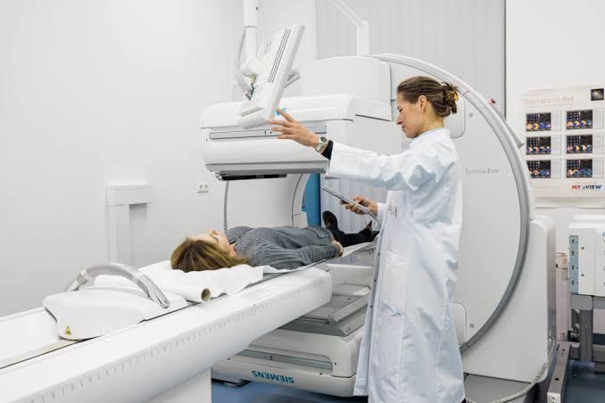
Renal imaging plays a crucial role in diagnosing and managing various kidney conditions in pediatric patients. While ultrasound and MRI provide detailed anatomical information, nuclear medicine renal scans offer unique insights into kidney function, blood flow, and drainage. These functional assessments are particularly valuable in children, where congenital anomalies, urinary tract infections, and obstructive conditions are common.
Understanding the Basics of Nuclear Medicine Renal Scans
Nuclear medicine imaging involves introducing a small, safe amount of a radioactive substance (radiopharmaceutical or tracer) into the body, typically via injection. This substance is designed to target specific organs or tissues. A specialized camera, called a gamma camera, detects the gamma rays emitted by the tracer as it moves through the body. A computer then processes this data to create images and graphs that depict organ function over time.
- Radiopharmaceutical: A drug tagged with a radioactive isotope. The choice of radiopharmaceutical depends on the specific function being assessed.
- Gamma Camera: Detects gamma radiation and converts it into an electrical signal, which is then processed into an image.
- Images/Data: Provide information about tracer distribution, uptake, transit, and excretion, offering functional insights not typically available from anatomical imaging techniques.
Static Renal Scans: The Technique and Pedatric Indications
Static renal scans, also known as DMSA scans, are primarily used to evaluate the structure and integrity of the renal parenchyma (the functional tissue of the kidney, specifically the cortex). They provide a static image of the kidneys after the radiopharmaceutical has accumulated in the tissue.
- Key Radiopharmaceutical: Technetium-99m (Tc-99m) Dimercaptosuccinic Acid (DMSA).
- Why DMSA? DMSA is a tracer that binds specifically to the tubules of the renal cortex. It is cleared very slowly from the kidney tissue, allowing for imaging at a later time point when the tracer distribution primarily reflects the mass of functioning cortical tissue.
- The Technique (Step-by-Step Understanding):
- 2a. Preparation:
- Parents/guardians receive clear instructions about the procedure.
- Adequate hydration is important, though typically less critical than for dynamic scans.
- For younger or uncooperative children, sedation may be necessary to ensure the child remains still during imaging.
- The child should be in a comfortable position, often lying down.
- 2b. Radiopharmaceutical Administration:
- A small dose of Tc-99m DMSA, calculated based on the child’s weight, is administered intravenously (usually through a vein in the arm or hand).
- 2c. Waiting Period:
- This is a crucial difference from dynamic scans. Imaging is performed 2-4 hours after injection (sometimes longer, up to 6 hours or 24 hours in specific cases, depending on institutional protocol and clinical question). This waiting period allows sufficient time for the DMSA to accumulate in the renal cortex and for background activity in the blood to decrease.
- 2d. Imaging Acquisition:
- The child is positioned under the gamma camera.
- Multiple views are acquired to visualize the kidneys from different angles:
- Posterior view (most common for initial assessment).
- Anterior view.
- Oblique views (posterior oblique left and right) – essential for separating the kidneys from overlying or underlying structures and better visualizing the kidney contours.
- Single-photon emission computed tomography (SPECT) may also be performed to create 3D images, improving the detection of subtle cortical defects.
- Images typically take several minutes per view. The child must remain very still.
- 2e. Image Interpretation:
- Images are assessed for:
- Size, shape, and location of the kidneys.
- Uniformity of radiotracer uptake throughout the cortex.
- Presence of focal or diffuse areas of decreased uptake, which represent areas of reduced or absent functioning cortical tissue. These defects are typically interpreted as areas of scarring (chronic damage) or inflammation (acute pyelonephritis).
- Split renal function can be estimated based on the relative uptake of DMSA in each kidney (see Section 4).
- Images are assessed for:
- 2a. Preparation:
- Pediatric Indications for Static (DMSA) Scan:
- Evaluation of Acute Pyelonephritis:
- DMSA scans are considered the most sensitive imaging modality for detecting acute pyelonephritis (kidney infection involving the renal parenchyma).
- Infection causes inflammation and temporary or permanent damage to cortical tissue, appearing as areas of decreased DMSA uptake.
- While not always necessary for diagnosing acute pyelonephritis based on clinical grounds, a DMSA scan can confirm renal parenchymal involvement, particularly in complex cases or when the diagnosis is uncertain.
- Identification of Renal Scarring following Urinary Tract Infection (UTI), especially with VUR:
- The primary and most common indication for DMSA scan in pediatrics is to detect permanent renal scarring (which appears as persistent cortical defects) that may result from pyelonephritis, especially in children with Vesicoureteral Reflux (VUR).
- VUR, the backward flow of urine from the bladder into the ureters and sometimes the kidneys, increases the risk of pyelonephritis. Recurrent pyelonephritis, particularly in the presence of high-grade VUR, can lead to irreversible renal scarring.
- Renal scarring can impair kidney growth and function and is a risk factor for hypertension and chronic kidney disease later in life.
- A DMSA scan performed typically 4-6 months after an episode of pyelonephritis is the standard method for assessing whether temporary inflammatory lesions have resolved or progressed to permanent scarring.
- Assessment of Renal Size and Shape:
- Provides functional images that complement anatomical imaging (like ultrasound) in evaluating congenital abnormalities in kidney size, shape, or position (e.g., horseshoe kidney, duplex kidney, ectopic kidney).
- Evaluation of Acute Pyelonephritis:
Dynamic Renal Scans: The Technique and Pediatric Indications
Dynamic renal scans, often performed with tracers like DTPA or MAG3, evaluate the blood supply to the kidneys, how the kidneys filter waste products from the blood, and how urine drains from the kidneys into the bladder. They provide a series of images acquired rapidly over time.
- Key Radiopharmaceuticals:
- Technetium-99m (Tc-99m) Diethylenetriaminepentaacetic Acid (DTPA): Filtered by the glomeruli (like GFR). Primarily evaluates glomerular filtration and transit.
- Technetium-99m (Tc-99m) Mercaptoacetyltriglycine (MAG3): Excreted by the renal tubules (like ERPF – Effective Renal Plasma Flow). More actively secreted, leading to higher kidney uptake, especially useful in patients with impaired renal function. Provides better image quality in children.
- The Technique (Step-by-Step Understanding):
- 3a. Preparation:
- Excellent hydration is critical. Children are encouraged to drink fluids before the scan to ensure adequate urine production and allow for effective assessment of drainage.
- The child should empty their bladder just before the study begins.
- An intravenous line is inserted for tracer injection and, if needed, diuretic administration.
- Sedation may be required for young or uncooperative children to ensure they remain still for the ~20-30 minute acquisition period.
- 3b. Radiopharmaceutical Administration and Dynamic Imaging:
- The gamma camera is positioned over the child’s abdomen and back, usually focusing on the kidneys and bladder region.
- The tracer (DTPA or MAG3, dose based on weight) is injected intravenously as a bolus (a rapid injection).
- Immediately after injection, the camera begins acquiring images continuously or in rapid sequence (e.g., every few seconds) for approximately 20-30 minutes. This captures the entire process: blood flow to the kidneys, uptake by the renal tissue, transit through the tubules, and drainage into the collecting system and bladder.
- 3c. Diuretic Administration (Diuretic Renogram):
- If the scan is specifically to evaluate hydronephrosis (swelling of the kidney due to impaired drainage), a diuretic (usually Furosemide, e.g., Lasix) is administered intravenously during the scan.
- The timing varies, often after the initial uptake phase (e.g., 15-20 minutes after tracer injection).
- The diuretic increases urine production, challenging the kidney’s drainage system. If a blockage exists, the increased urine volume will build up above the obstruction.
- 3d. Data Processing and Analysis:
- The sequence of images is processed by a computer.
- Regions of interest (ROIs) are drawn around each kidney and the bladder.
- Time-Activity Curves: The computer generates graphs showing the amount of tracer in each ROI over time. These curves are the primary tools for interpreting dynamic renal scans:
- Perfusion Phase: A rapid rise corresponding to blood flow to the kidney.
- Cortical Uptake/Transit Phase: Tracer enters and moves through the renal parenchyma.
- Excretion Phase: Tracer moves from the renal pelvis down the ureter into the bladder. In a non-obstructed system, the curve for the kidney ROI will show a decline after reaching a peak, especially after diuretic administration. In an obstructed system, the curve will remain flat or continue to rise, indicating urine pooling.
- Split Renal Function: Calculated precisely from the count data in the kidney ROIs over a specific time interval (see Section 4).
- 3a. Preparation:
- Pediatric Indications for Dynamic Renal (DTPA/MAG3) Scan:
- Evaluation of Hydronephrosis:
- This is a major indication. Hydronephrosis, often detected on prenatal ultrasound, refers to dilation of the renal pelvis and calyces. The dynamic scan, particularly the diuretic renogram, helps determine if the dilation is due to a significant obstruction impairing urine flow or if it’s a non-obstructive dilation (“pelviectasis”) that doesn’t impede drainage.
- The pattern of the time-activity curve after diuretic challenge indicates the severity of any obstruction and helps predict whether surgical intervention might be necessary.
- Assessment of Renal Function:
- Provides quantitative estimates of overall and individual kidney function (glomerular filtration rate with DTPA, effective renal plasma flow with MAG3).
- Useful for monitoring function in various medical renal diseases.
- Evaluation of Renal Blood Flow:
- The initial phase of the scan provides information about perfusion to each kidney.
- Evaluation of Renal Transplants:
- Used to assess initial engraftment, perfusion, and function, and to investigate potential complications like vascular compromise or urinary obstruction.
- Evaluation of Hydronephrosis:
The Concept of Split Renal Function
Split renal function, also known as differential renal function, refers to the percentage contribution of each individual kidney to the total clearance function of both kidneys combined.
- What it is: If the total clearance is 100%, split function tells you, for example, if the right kidney contributes 55% and the left contributes 45%.
- How it’s Determined:
- Static (DMSA) Scan: Estimates split function based on the relative amount of DMSA uptake in each kidney after the waiting period, assuming DMSA uptake is proportional to the amount of functioning cortical tissue.
- Dynamic (DTPA/MAG3) Scan: Calculates split function more precisely by measuring the percentage of tracer taken up by each kidney from the bloodstream during the initial uptake phase (before significant excretion occurs). MAG3 is often preferred for split function calculation, especially in kidneys with impaired function, as its secretion mechanism leads to higher uptake relative to glomerular filtration.
- Why it’s Important in Pediatrics:
- Tracking Disease Progression: Allows monitoring of changes in function of an affected kidney over time (e.g., in VUR, pyelonephritis, or congenital anomalies).
- Diagnosis and Management: Provides crucial information for conditions like unilateral kidney disease or differences in function between kidneys.
- Surgical Planning: Essential for planning surgeries that affect kidney tissue (e.g., partial nephrectomy) or the urinary tract (e.g., anti-reflux surgery, pyeloplasty for hydronephrosis). Knowing the function of the potentially compromised kidney and the contralateral kidney helps determine risk and prognosis.
- Transplant Evaluation: Assesses the function of the transplanted kidney and the native kidneys (if still present).
Normal split function is typically close to 50/50, although minor variations (e.g., 52/48) are common. Significant deviations from symmetry (e.g., one kidney contributing less than 40%) can indicate reduced function in that kidney, potentially due to scarring, obstruction, or agenesis/dysplasia.
Pediatric-Specific Considerations
Performing renal scans in children requires specific attention to:
- Patient Cooperation: Sedation protocols and child-friendly environments are often necessary.
- Hydration: Ensuring proper hydration is crucial for both types of scans, but particularly for dynamic drainage studies.
- Radiation Dose: Doses of radiopharmaceuticals are carefully calculated based on weight or body surface area following the ALARA principle (As Low As Reasonably Achievable).
- Parental Support: Educating parents about the procedure and allowing them to be present (following safety guidelines) can help ease the child’s anxiety.
Conclusion
Static (DMSA) and dynamic (DTPA/MAG3) renal scans are indispensable tools in pediatric nephrology and urology, providing functional information that complements anatomical imaging.
- Static (DMSA) scans are the gold standard for visualizing renal cortical parenchyma and detecting scarring following pyelonephritis, especially in the context of VUR.
- Dynamic (DTPA/MAG3) scans, particularly with diuretic challenge, are essential for evaluating kidney blood flow, precise function, and determining the significance of upper urinary tract dilation like hydronephrosis by assessing drainage.
- The concept of split renal function, calculable from both scan types (more accurately with dynamic scans), is vital for monitoring kidney health, informing clinical decisions, and guiding surgical interventions in children with congenital or acquired renal conditions.
These techniques, performed with careful attention to pediatric needs, provide critical data for optimizing the diagnosis, management, and long-term outcomes for young patients with kidney and urinary tract disorders.


