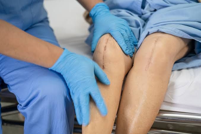
Understanding Wound Healing: A Comprehensive Overview
Wound healing is a complex, dynamic biological process crucial for restoring tissue integrity and function following injury. It involves a coordinated cascade of cellular and molecular events. A thorough understanding of this process, its influencing factors, and associated clinical considerations is essential for effective wound management. This document outlines fundamental concepts related to wounds and their healing, presented in a clear, structured format.
Defining a Wound
A wound is medically defined as a disruption of the normal structure and function of tissue, typically skin, caused by trauma, surgery, or underlying pathology. Wounds can range from minor abrasions and lacerations to deep incisions, burns, ulcers, or crush injuries. They represent a break in the body’s protective barrier, potentially exposing underlying tissues to infection and impairing physiological functions. The type, depth, and extent of a wound significantly influence the healing process.
Sequence and Approximate Time Frame of the Phases of Wound Healing
Wound healing proceeds through a predictable, overlapping sequence of four main phases. The exact timing can vary based on the wound type, location, and the patient’s overall health, but a general time frame can be outlined:
-
- Phase 1: Hemostasis Phase (Immediately after injury)
- Description: This is the initial and shortest phase, occurring immediately upon injury. Its primary goal is to stop bleeding.
- Key Events: Vasoconstriction to reduce blood flow; platelet aggregation to form a plug; activation of the coagulation cascade, resulting in the formation of a fibrin clot, which acts as a temporary matrix and provides scaffolds for migrating cells.
- Approximate Time Frame: Minutes to hours.
- Phase 2: Inflammatory Phase (Hours to days after injury)
- Description: Also known as the defensive phase, this phase is initiated by the injury itself and the presence of necrotic tissue and pathogens. It aims to clear the wound site of debris, bacteria, and damaged tissue and prepare it for subsequent repair.
- Key Events: Vasodilation and increased capillary permeability (leading to redness, swelling, heat, pain); infiltration of leukocytes (neutrophils arrive first to phagocytose bacteria and debris, followed by macrophages, which continue phagocytosis, release growth factors, and transition the wound to the next phase); release of inflammatory mediators (cytokines, chemokines).
- Approximate Time Frame: Peaks around 24-48 hours and typically lasts 4-6 days. Chronic inflammation can significantly impede healing.
- Phase 3: Proliferative Phase (Days to weeks after injury)
- Description: This phase focuses on rebuilding the injured tissue. It involves the formation of new tissue components and covering the wound surface.
- Key Events:
- Angiogenesis: Formation of new blood vessels from pre-existing ones to supply nutrients and oxygen to the healing tissue.
- Granulation Tissue Formation: Development of a rich, vascular connective tissue (discussed in detail below).
- Collagen Deposition: Fibroblasts migrate into the wound and synthesize collagen (primarily Type III initially), which forms the structural matrix of the new tissue.
- Epithelialization: Epithelial cells migrate from the wound edges across the granulation tissue to cover and close the wound surface.
- Wound Contraction: Myofibroblasts in the granulation tissue contract, pulling the wound edges together, reducing the wound size.
- Approximate Time Frame: Commences around day 4-6 and can last for several weeks, depending on wound size and depth.
- Phase 4: Maturation (Remodeling) Phase (Weeks to years after injury)
- Description: This is the final, longest phase during which the newly formed tissue is strengthened and reorganized to resemble the original tissue as closely as possible.
- Key Events: Collagen fibers are reorganized, cross-linked, and remodeled from Type III to stronger Type I collagen; scar tissue becomes stronger, less vascular (paling in color), and more pliable; cellularity decreases.
- Approximate Time Frame: Can begin around week 3 and continue for months or even years. Scar tissue typically achieves about 80% of the original tissue’s tensile strength.
- Phase 1: Hemostasis Phase (Immediately after injury)
Essential Elements and Significance of Granulation Tissue
Granulation tissue is a hallmark of the proliferative phase of healing, particularly in wounds healing by secondary intention. It is a temporary, highly vascular connective tissue that fills the wound defect.
-
- Essential Elements: Granulation tissue is primarily composed of:
- Fibroblasts: Cells responsible for synthesizing the extracellular matrix (especially collagen, elastin, and proteoglycans).
- New capillaries (angiogenesis): Small, delicate blood vessels that give the tissue a characteristic pink or red, granular appearance and supply essential nutrients, oxygen, and immune cells. Endothelial cells form these vessels.
- Macrophages: Continue to clean debris and release growth factors that stimulate fibroblast and endothelial cell activity.
- Extracellular Matrix: Primarily collagen laid down by fibroblasts, providing structural support.
- Significance: Granulation tissue is critically important because it:
- Provides a scaffold for the migration of fibroblasts and endothelial cells.
- Fills the wound void, reducing dead space.
- Serves as a bed for epithelial cell migration during epithelialization.
- Contains myofibroblasts, which drive wound contraction.
- Protects the underlying tissue from infection (though fragile).
- Without sufficient, healthy granulation tissue, a wound cannot successfully progress through the proliferative and maturation phases, leading to delayed or impaired healing.
- Essential Elements: Granulation tissue is primarily composed of:
Types of Wound Healing and the Elements of Each
Wounds heal by different “intentions” based on the amount of tissue loss, the presence of infection, and whether the wound edges are approximated.
-
- Primary Intention (Primary Closure):
- Description: Occurs when wound edges are clean, straight, and closely approximated, typically through surgical closure (sutures, staples, adhesive). Minimal tissue loss and dead space are present.
- Elements: Inflammation is minimal; epithelialization occurs rapidly across the narrow gap; granulation tissue formation and wound contraction are minimal. Scarring is typically fine (linear scar).
- Examples: Surgically closed incisions, clean lacerations closed promptly.
- Secondary Intention (Spontaneous Healing):
- Description: Occurs in wounds with significant tissue loss, irregular edges, or contamination/infection. The wound is left open and allowed to heal from the bottom up.
- Elements: Inflammation is more intense and prolonged compared to primary intention; significant granulation tissue formation is required to fill the large defect; substantial wound contraction occurs to reduce the wound size; epithelialization occurs slowly, migrating inward from the periphery over the large area of granulation tissue. Scarring is typically more prominent and extensive.
- Examples: Pressure ulcers, large burns, traumatic wounds with tissue loss, infected wounds left open.
- Tertiary Intention (Delayed Primary Closure):
- Description: Occurs when a wound is initially left open (like secondary intention) due to contamination or swelling, but is later closed surgically once the infection risk is reduced or swelling subsides. It combines elements of both primary and secondary healing.
- Elements: Initial phase involves debridement and management to reduce contamination (similar to initial secondary healing); if clean and healthy granulation tissue forms, the wound is then surgically closed (similar to primary healing). This results in less granulation/contraction than secondary healing but more than primary healing, with a wider scar than primary closure.
- Examples: Contaminated traumatic wounds, appendectomy incision where infection is suspected.
- Primary Intention (Primary Closure):
Phases of Wound Healing Distinct to Each Type of Wound
While the sequence of phases (Hemostasis, Inflammation, Proliferation, Maturation) is universal, their duration and intensity differ significantly depending on the type of healing intention.
-
- Primary Intention:
- Inflammatory Phase: Brief and low intensity due to minimal tissue damage and contamination.
- Proliferative Phase: Focus is primarily on rapid epithelialization across the narrow gap and minimal collagen deposition to bind the edges. Granulation tissue formation is minimal, confined to the narrow space. Wound contraction is negligible.
- Maturation Phase: Involves remodeling of the small amount of collagen laid down, resulting in a fine, linear scar.
- Secondary Intention:
- Inflammatory Phase: More prolonged and intense due to greater tissue damage, debris, and potential contamination. Essential for clearing the large wound bed.
- Proliferative Phase: This phase is extensive and crucial. It involves significant and sustained angiogenesis and robust granulation tissue formation to fill the substantial tissue defect. Wound contraction by myofibroblasts plays a major role in reducing wound size. Epithelialization is slow, covering the large new surface from the periphery.
- Maturation Phase: Extended and involves significant remodeling of the large volume of granulation tissue/collagen laid down, resulting in a larger, often more complex scar that may contract significantly.
- Tertiary Intention:
- Initial Phase: Follows the inflammatory phase with a period resembling the early proliferative phase of secondary healing, allowing granulation tissue to form in an open wound.
- Delayed Closure: Once closed, the healing mechanism transitions towards that of primary intention. The inflammatory phase, if prolonged initially, subsides. Subsequent proliferation and maturation proceed more like primary intention but build upon the granulation tissue already formed. Contraction is less than secondary but more than primary.
- Primary Intention:
Clinical Factors That Decrease Collagen Synthesis and Retard Wound Healing
Collagen synthesis is vital for the structural integrity of new tissue. Numerous factors can impair this process and delay or halt wound healing:
-
- Nutritional Deficiencies: Protein (building blocks for collagen), Vitamin C (essential cofactor for collagen synthesis), Vitamin A (stimulates epithelialization and collagen synthesis), Zinc (cofactor for enzymes involved in collagen synthesis and cell proliferation).
- Poor Blood Supply/Ischemia: Reduced delivery of oxygen and nutrients, which are critical for cellular metabolism and collagen production. Conditions like peripheral vascular disease, diabetes, and pressure on tissues contribute.
- Infection: Bacteria consume nutrients, produce toxins, and release enzymes that degrade collagen and other matrix components, diverting resources from repair to fighting infection.
- Corticosteroids: Systemic or topical corticosteroids inhibit inflammation, reduce fibroblast proliferation, and impair collagen synthesis and wound contraction.
- Diabetes Mellitus: Poorly controlled diabetes affects multiple healing phases: impaired neutrophil function, reduced angiogenesis, decreased fibroblast function and collagen synthesis, neuropathy affecting sensation, and vascular complications leading to ischemia.
- Smoking: Nicotine causes vasoconstriction, reducing tissue perfusion and oxygen delivery. Carbon monoxide reduces oxygen-carrying capacity. Smoking impairs fibroblast function and collagen synthesis.
- Advanced Age: While not necessarily preventing healing, the process is generally slower in older individuals due to reduced cellular proliferation, decreased inflammatory response, and comorbidities.
- Chronic Disease States: Conditions like renal failure, liver disease, and immunosuppression can impair metabolic processes and immune function necessary for healing.
- Mechanical Stress/Pressure: Repeated trauma or pressure on the wound site disrupts the delicate healing tissue and blood vessels, hindering collagen deposition and leading to breakdown.
- Radiation Therapy: Can damage fibroblasts and blood vessels, leading to chronic non-healing wounds or tissue fibrosis (excessive, disorganized collagen).
Rationale for the Uses of Absorbable and Nonabsorbable Sutures
Sutures are used to approximate tissue edges and support the wound during the early healing phases. The choice between absorbable and nonabsorbable sutures depends on the tissue being repaired and the expected duration of support required.
-
- Absorbable Sutures:
- Rationale: These sutures are designed to be broken down and absorbed by the body’s enzymatic hydrolysis or phagocytosis over a specific time frame (weeks to months). They provide temporary support until the tissue has gained sufficient tensile strength through collagen deposition.
- Uses: Used for internal tissues that heal relatively quickly and do not require permanent support (e.g., subcutaneous tissue, fascia, muscle, ligating blood vessels) or in locations where suture removal would be difficult or unnecessary (e.g., deep layers of a surgical incision, gynecology, urology).
- Nonabsorbable Sutures:
- Rationale: These sutures are made of materials that resist breakdown by the body and provide permanent or prolonged support.
- Uses: Used where long-term or permanent tissue approximation is needed or in tissues that heal very slowly and require extended support. This includes skin closure (requiring later removal), vascular anastomosis, tendon repairs, fascia in areas under tension, or prosthetic material fixation. They are left in place indefinitely (if internal) or removed after the wound has healed sufficiently (if external).
- Absorbable Sutures:
Functions of a Dressing
Wound dressings play multiple critical roles in optimizing the healing environment and protecting the wound. Key functions include:
-
- Protection: Shielding the wound from mechanical trauma, contamination (bacteria, foreign bodies), and infection.
- Moisture Balance: Maintaining a physiologically moist wound environment, which is crucial for cell migration (epithelialization, fibroblast activity) and the function of growth factors and enzymes. Avoiding excessive dryness (impedes cell movement) and excessive wetness (causes maceration of surrounding skin).
- Absorption: Managing wound exudate (fluid) to prevent maceration of the wound edges and surrounding skin, while retaining essential moisture.
- Thermal Insulation: Maintaining a stable temperature at the wound surface, as healing proceeds optimally within a narrow temperature range.
- Pain Management: Protecting nerve endings from exposure and mechanical irritation, and some dressings containing analgesic properties.
- Debridement: Some dressings (e.g., hydrogels, enzymatic dressings) can assist in autolytic or enzymatic debridement of necrotic tissue.
- Homeostasis: Some dressings (e.g., alginates, foams) have hemostatic properties to help control minor bleeding.
- Reducing Edema: Compression dressings can help reduce swelling.
- Delivery of Topical Agents: Dressings can hold and deliver medications or other therapeutic agents to the wound bed.
Defining Clean, Contaminated, and Infected Wounds and Describing the Management of Each
Classifying wounds based on their bacterial load is crucial for guiding management strategies and predicting healing outcomes.
-
- Clean Wound:
- Definition: An uninfected operative wound in which no inflammation is encountered and the respiratory, alimentary, genital, or uninfected urinary tract is not entered. Primarily closed.
- Management: Closure by primary intention is the standard. Strict aseptic technique is paramount to prevent contamination. Postoperative care focuses on keeping the wound clean and dry and monitoring for signs of infection (rare if properly managed).
- Contaminated Wound:
- Definition: An open, fresh, accidental wound (e.g., from trauma) or an operative wound involving a major break in sterile technique or gross spillage from the gastrointestinal tract, or incisions encountering acute, nonpurulent inflammation. Contains a significant bacterial burden but not actively infected (typically 10^2 – 10^5 bacteria per gram of tissue).
- Management: Closure by primary intention is generally avoided unless the wound is thoroughly debrided and irrigated. Often managed initially by thorough cleaning, copious irrigation, and debridement of devitalized tissue. May be left open to heal by secondary intention or managed with delayed primary closure (tertiary intention) after a period of observation and local wound care to ensure no infection develops. Prophylactic antibiotics may be considered depending on the context.
- Infected Wound:
- Definition: A wound where microorganisms are actively proliferating and causing signs of infection, such as increased pain, redness (erythema), swelling (edema), warmth, pus (purulent exudate), odor, and potentially systemic signs like fever. Bacterial load typically exceeds 10^5 organisms per gram of tissue. Can originate from trauma, surgery, or contamination of a chronic wound.
- Management: Surgical management is focused on controlling the infection:
- Opening the wound: If closed, the wound must be opened to allow drainage of pus and exudate.
- Debridement: Extensive debridement of necrotic and infected tissue is essential.
- Irrigation: Copious irrigation to reduce bacterial load.
- Antibiotics: Systemic antibiotics are necessary, chosen based on suspected pathogens or culture and sensitivity results.
- Packing/Drainage: The wound is left open and often packed to allow continued drainage and healing by secondary intention. Topical antimicrobials may also be used.
- Once the infection is controlled and a clean wound bed with healthy granulation tissue is established, the wound will heal by secondary intention or may be considered for delayed closure or grafting.
- Clean Wound:
In conclusion, effective wound management requires not only a deep understanding of the intricate biological phases of healing but also the ability to assess the wound’s characteristics, classify its type, identify factors that may impede progress, and select appropriate interventions, such as sutures and dressings, to support the body’s natural regenerative capabilities.


