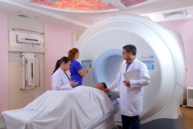
Introduction to Nuclear Medicine: A Guide for Medical Students
Welcome to an introduction to the fascinating field of nuclear medicine, a medical specialty that uses small amounts of radioactive materials to diagnose and treat a variety of diseases. Unlike conventional anatomical imaging modalities like X-ray, CT, or MRI, nuclear medicine focuses on revealing physiological function and metabolic activity, often detecting diseases in their earliest stages. This guide aims to provide you with a foundational understanding of this unique discipline.
Understanding the Core Concept
At its heart, nuclear medicine is about tracing biochemical processes within the body. It involves:
- Introducing a Radiopharmaceutical: This is a compound formed by attaching a radioactive atom (an isotope, or “radioisotope”) to a pharmaceutical molecule. The pharmaceutical is chosen because it targets a specific organ, tissue, or cellular process (e.g., glucose metabolism, bone turnover, blood flow).
- Distribution within the Body: Once administered (usually intravenously, but sometimes orally or by inhalation), the radiopharmaceutical travels and accumulates in the target area based on the body’s physiological function.
- Detection of Gamma or Positron Emission: The radioisotope decays, emitting detectable particles or energy (typically gamma rays or positrons).
- Image Formation: specialized cameras (like gamma cameras or PET scanners) detect this emitted radiation. A computer then processes the data to create images that show the spatial distribution and concentration of the radiopharmaceutical within the body. Areas of high uptake appear “hot,” while areas of low uptake appear “cold” or less intense.
Key Takeaway: While other imaging modalities show you what structures are present (anatomy), nuclear medicine shows you how those structures and the body as a whole are functioning (physiology and metabolism).
The Building Blocks – Radiopharmaceuticals and Imaging Equipment
Understanding the components is crucial:
- Radiopharmaceuticals (Radiotracers): These are the stars of the show. The choice of radiopharmaceutical depends entirely on the physiological process or pathology being investigated.
- Common Radioisotopes:
- Technetium-99m (Tc-99m): The most widely used isotope due to its ideal physical properties (6-hour half-life, 140 keV gamma ray). It can be attached to various pharmaceuticals for imaging bones, heart, kidneys, brain, liver, and more.
- Fluorine-18 (F-18): Primarily used in Positron Emission Tomography (PET). F-18 Fluorodeoxyglucose (FDG) is the most common, tracing glucose metabolism, which is elevated in many cancers and inflamed tissues.
- Iodine-123 (I-123): Used for thyroid imaging and uptake studies.
- Iodine-131 (I-131): Used for larger dose thyroid uptake studies and, importantly, for treatment of thyroid diseases (hyperthyroidism, thyroid cancer).
- Thallium-201 (Tl-201): Used for myocardial perfusion imaging.
- The Pharmaceutical: This component determines where the tracer goes. Examples include Methylene Diphosphonate (MDP) which targets bone turnover, Sestamibi or Myoview which target viable myocardial cells, or FDG which acts as a glucose analogue.
- Common Radioisotopes:
- Imaging Equipment:
- Gamma Camera (SPECT): Single Photon Emission Computed Tomography. This camera has a crystal that scintillates (emits light) when struck by gamma rays. Photomultiplier tubes detect the light, and a computer maps the location of emissions. By rotating the camera around the patient, 3D images (SPECT) can be reconstructed, providing better lesion localization than planar images.
- PET Scanner (PET/CT, PET/MRI): Positron Emission Tomography. PET isotopes emit positrons, which travel a short distance before annihilating with an electron, producing two gamma rays traveling in opposite directions (180 degrees apart). The PET scanner detects these coincident gamma rays. PET is generally more sensitive and provides higher spatial resolution than SPECT. Modern PET scanners are often combined with CT or MRI scanners (PET/CT, PET/MRI) to provide combined functional and anatomical information on a single set of images.
Radiation Safety Principles
Medical students should be aware of the basic principles of radiation safety:
- ALARA: As Low As Reasonably Achievable. This principle guides all nuclear medicine procedures to minimize radiation dose to patients, staff, and the public.
- Time, Distance, Shielding: Minimizing time spent near radioactive sources, maximizing distance from the source, and using appropriate shielding (e.g., lead) are fundamental safety measures.
- Patient Instructions: Patients receiving radiopharmaceuticals are often given specific instructions regarding limiting close contact with others (especially children and pregnant women) for a period after the scan, as they temporarily emit radiation.
Common Nuclear Medicine Applications and Tests
Nuclear medicine is applied across many medical specialties. Here are some common examples:
- Oncology: PET/CT with F-18 FDG is widely used for cancer staging, restaging, treatment response assessment, and detecting recurrence for many tumor types (lung, lymphoma, melanoma, colorectal, head and neck, etc.). Other tracers target specific tumor types (e.g., Ga-68 DOTATATE for neuroendocrine tumors, PSMA PET for prostate cancer).
- Cardiology: Myocardial Perfusion Imaging (MPI) assesses blood flow to the heart muscle at rest and during stress to detect coronary artery disease (ischemia or infarction). Viability studies (using FDG PET or Thallium) assess if scarred myocardium is still alive and potentially recoverable with revascularization.
- Skeletal System: Bone Scans (Tc-99m MDP) are highly sensitive for detecting increased bone metabolism, commonly used for identifying bone metastases, stress fractures, infections (osteomyelitis), or inflammatory processes (arthritis).
- Endocrinology: Thyroid scans and uptake studies evaluate thyroid function, size, shape, and nodules (I-123, Tc-99m). I-131 is used for treating hyperthyroidism and differentiated thyroid cancer. MIBG scans (I-123 or I-131) are used for neuroendocrine tumors like pheochromocytomas.
- Nephrology: Renal scans (Tc-99m MAG3, DTPA) assess kidney perfusion, function (glomerular filtration, tubular secretion), and obstruction.
- Gastroenterology: HIDA scans (Tc-99m iminodiacetic acid derivatives) evaluate gallbladder function and patency of the biliary system (cystic duct obstruction, acute cholecystitis). Gastric emptying scans assess the rate food leaves the stomach.
- Neurology: Brain perfusion SPECT (Tc-99m HMPAO/ECD) can evaluate regional cerebral blood flow patterns in stroke, dementia, or seizures. Amyloid PET scans (e.g., F-18 Florbetapir) help diagnose Alzheimer’s disease by visualizing amyloid plaques.
- Infection/Inflammation: Scans using gallium-67 or labeled white blood cells (Indium-111 or Tc-99m HMPAO-labeled leukocytes) can help localize occult infections or inflammatory conditions.
Interpreting Images – Normal vs. Pathological Examples
Interpreting nuclear medicine images involves understanding the expected biodistribution of the radiopharmaceutical in normal physiology and recognizing deviations that indicate pathology. Here are descriptive examples:
- Example 1: Tc-99m MDP Bone Scan
- Purpose: Evaluate bone metabolism, primarily for detecting metastases and fractures.
- Normal Appearance: Symmetrical, relatively uniform uptake throughout the adult skeleton. Areas of normally higher uptake include the sacroiliac joints, sternum, costochondral junctions, shoulders, and knees (due to higher metabolic activity or superimposed structures). The kidneys and bladder are also visible due to tracer excretion.
- Pathological Appearance: The most common abnormality is a focal area of increased uptake (“hot spot”). A single hot spot could be a benign lesion or fracture. Multiple hot spots distributed randomly throughout the axial and appendicular skeleton are highly suspicious for bone metastases. Diffuse increased uptake in specific patterns can suggest metabolic bone disease or widespread metastases. A focal area of decreased uptake (“cold spot”) is less common but can indicate avascular necrosis, myeloma, or certain aggressive bone tumors where bone destruction outpaces reactive uptake.
- Example 2: Stress/Rest Myocardial Perfusion Scan (e.g., using Tc-99m Sestamibi)
- Purpose: Assess blood flow to the heart muscle to detect ischemia or infarction. Images are acquired after physical or pharmacological stress and again after rest.
- Normal Appearance: Homogeneous (uniform) uptake of the radiotracer throughout the wall of the left ventricle in both stress and rest images. This indicates adequate blood flow to all regions of the myocardium under both conditions.
- Pathological Appearance:
- Reversible Defect (Ischemia): Reduced radiotracer uptake in a specific region of the left ventricle on the stress images that fills in (shows increased uptake) on the rest images. This indicates reduced blood flow only under stress, consistent with ischemia due to a flow-limiting coronary stenosis.
- Fixed Defect (Infarction/Scar): Reduced radiotracer uptake in a specific region of the left ventricle on both the stress and rest images. This indicates a permanent loss of viable myocardial tissue (infarct or scar) in that region.
- The location of these defects corresponds to the territory supplied by specific coronary arteries.
- Example 3: F-18 FDG PET/CT Scan
- Purpose: Assess glucose metabolism, primarily for cancer detection, staging, and monitoring response to treatment.
- Normal Appearance: High FDG uptake is physiologically normal in areas with high glucose metabolism: the brain, heart (variable depending on fasting state), kidneys and bladder (tracer excretion), sometimes liver, spleen, and bone marrow. Physiologic uptake can also be seen in activated muscles (if recent exercise), brown fat (especially in cold environments, typically in the neck and upper chest), and lymphoid tissue (tonsils, thymus in children, reactive lymph nodes). The CT provides anatomical context.
- Pathological Appearance: Focal areas of abnormally increased FDG uptake, significantly higher than background or surrounding healthy tissue, that do not correspond to normal physiological sites. In the context of oncology, such findings are highly suspicious for malignant tumors, as many cancers exhibit increased glucose metabolism (Warburg effect). However, increased FDG uptake can also be seen in inflammatory or infectious processes, which are also metabolically active. The CT component helps distinguish between these possibilities and precisely localize the metabolic abnormality to an anatomical structure (e.g., a suspicious lung nodule, an enlarged lymph node).
- Example 4: I-123 Thyroid Scan and Uptake
- Purpose: Evaluate thyroid size, shape, location, and function.
- Normal Appearance: The scan shows homogeneous uptake of the radiotracer throughout both thyroid lobes, with the isthmus connecting them. The shape is typically a symmetrical “butterfly” appearance in the lower neck. A quantitative uptake measurement (radioiodine uptake, RAUI) falls within a normal range (dependent on dietary iodine intake, often 10-30% at 24 hours).
- Pathological Appearance:
- Nodules: Scans are crucial for evaluating thyroid nodules. A nodule with increased uptake (“hot nodule”) is often hyperfunctioning and usually benign. A nodule with decreased or absent uptake (“cold nodule”) is non-functioning and has a higher (though still low) probability of being malignant, requiring further investigation (e.g., fine needle aspiration biopsy).
- Diffuse Goiter: Enlarged gland with diffuse uptake.
- Graves’ Disease: Diffuse increased uptake throughout the gland, often with an enlarged appearance, and a high RAUI.
- Thyroiditis (e.g., Hashimoto’s): Often diffuse decreased uptake and a low RAUI (in the hypothyroid phase).
- Absent Uptake: Can indicate complete destruction of the gland or agenesis.
Advantages and Limitations of Nuclear Medicine
- Advantages:
- Functional Information: Provides unique physiological and metabolic insights not available from anatomical imaging alone.
- High Sensitivity: Can often detect disease processes at a molecular or cellular level before structural changes are evident on other scans.
- Whole-Body Imaging: Many scans (like bone scans, PET/CT) can image the entire body, valuable for detecting widespread disease like metastases or infection.
- Treatment Monitoring: Functional changes can often be assessed earlier than structural changes, aiding in monitoring treatment response.
- Therapeutic Applications: Some radioisotopes (like I-131, Lu-177) are used to treat diseases (theranostics).
- Limitations:
- Lower Spatial Resolution: Generally provides less detailed anatomical resolution compared to CT or MRI (though PET/CT and SPECT/CT help overcome this by fusing with anatomical images).
- Radiation Exposure: Patients receive a small radiation dose. Procedures must be justified and optimized (ALARA).
- Scan Time: Some scans can take longer than CT or MRI.
- Tracers Aren’t Always Specific: Increased uptake can sometimes be due to benign conditions (e.g., inflammation mimicking cancer on PET/CT).
- Availability and Cost: Access to certain tracers and equipment can be limited.
Integrating Nuclear Medicine into Clinical Practice
As a medical student, you will encounter nuclear medicine reports and images during rotations. Understanding the basic principles will help you:
- Interpret reports in the context of the patient’s history and other imaging.
- Appreciate the unique contribution nuclear medicine offers to diagnosis, staging, and treatment monitoring.
- Understand the indications and contraindications for ordering nuclear medicine tests.
- Communicate basic information to patients regarding their scan (e.g., preparation, duration, safety precautions).
Conclusion
Nuclear medicine is a dynamic and evolving field that plays a vital role in modern healthcare by providing unique functional and metabolic information. By understanding the fundamental principles of radiopharmaceuticals, imaging technologies, and image interpretation, you will be better equipped to utilize this powerful diagnostic and therapeutic tool throughout your medical career. Don’t hesitate to ask questions and engage with nuclear medicine professionals during your training to deepen your understanding.


