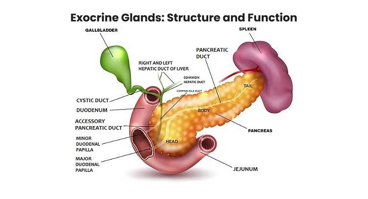
Differentiating Endocrine and Exocrine Glands
The primary distinction between endocrine and exocrine glands lies in the destination and mode of transport of their secretions.
- Exocrine Glands:
- Secrete substances into a duct system.
- These ducts carry the secretions to a specific location, typically an external surface of the body (like skin) or an internal surface that connects to the outside world (like the lining of the digestive tract, respiratory tract, or reproductive tract).
- Secretions are usually not hormones; they include enzymes (e.g., digestive enzymes from the pancreas), mucus (e.g., goblet cells in the respiratory tract), sweat (e.g., sweat glands in the skin), saliva (e.g., salivary glands), milk (e.g., mammary glands), and sebum (e.g., sebaceous glands).
- Their effects are typically localized to the site where the secretion is delivered.
- Examples: Salivary glands, sweat glands, mammary glands, glands in the lining of the stomach and intestines, pancreas (exocrine part secreting digestive enzymes), liver (secreting bile into ducts).
- Endocrine Glands:
- Are “ductless” glands.
- Secrete chemical messengers called hormones directly into the bloodstream or the interstitial fluid surrounding the cells.
- From the bloodstream, hormones travel throughout the body to reach target cells or target organs that have specific receptors for that hormone.
- Hormones exert their effects on these distant target cells, influencing their activity, growth, or metabolism.
- Their effects are typically widespread and systemic, although action is limited to cells with the appropriate receptors.
- Examples: Pituitary gland, thyroid gland, parathyroid glands, adrenal glands, pancreas (endocrine part secreting insulin and glucagon), gonads (ovaries and testes), pineal gland.
Here is a summary of the key differences:
| Feature | Exocrine Glands | Endocrine Glands |
|---|---|---|
| Presence of Ducts | Yes | No (Ductless) |
| Nature of Secretion | Enzymes, mucus, sweat, oils, milk, etc. | Hormones |
| Destination of Secretion | Body surface or lumen of a tract via ducts | Bloodstream or interstitial fluid |
| Mode of Transport | Via ducts | Via bloodstream or diffusion |
| Target | Local surface/lumen | Distant target cells/organs via blood |
| Speed of Action | Generally faster, localized | Generally slower, systemic, longer-lasting |
| Examples | Salivary glands, sweat glands, pancreas (exocrine), liver (bile) | Pituitary, Thyroid, Adrenal, Pancreas (endocrine), Gonads |
The Major Endocrine Glands
The human endocrine system comprises several dedicated endocrine glands, as well as organs that contain endocrine tissue as part of their function. The major endocrine glands are:
- Hypothalamus: Located in the brain, it produces releasing and inhibiting hormones that regulate the pituitary gland, and also produces ADH and oxytocin.
- Pituitary Gland (Hypophysis): A small gland located at the base of the brain, beneath the hypothalamus. It is often called the “master gland” because it regulates many other endocrine glands. It has two main parts: the anterior pituitary and the posterior pituitary, each producing different hormones.
- Pineal Gland: Located in the brain, it primarily produces melatonin, involved in regulating sleep-wake cycles.
- Thyroid Gland: Located in the neck, it produces thyroid hormones (T3 and T4), which regulate metabolism, and calcitonin, which helps regulate calcium levels.
- Parathyroid Glands: Usually four small glands located on the posterior surface of the thyroid gland. They produce parathyroid hormone (PTH), which is crucial for regulating calcium and phosphate levels in the blood.
- Adrenal Glands: Two glands located atop the kidneys. Each gland has two functionally distinct parts:
- Adrenal Cortex: Produces corticosteroids (e.g., cortisol, aldosterone).
- Adrenal Medulla: Produces catecholamines (epinephrine and norepinephrine).
- Pancreas (Endocrine Islets of Langerhans): Located behind the stomach, the pancreas has both exocrine (digestive enzymes) and endocrine functions. The endocrine function is carried out by the Islets of Langerhans, which produce insulin and glucagon, vital for blood glucose regulation.
- Gonads:
- Ovaries (in females): Located in the pelvic cavity, they produce estrogen and progesterone, involved in sexual development and reproduction.
- Testes (in males): Located in the scrotum, they produce testosterone, involved in sexual development and reproduction.
- Thymus: Located in the upper chest, it is more active in childhood. While primarily a lymphatic organ, it produces hormones like thymosin, which are important for the development of T lymphocytes.
Other organs also contain endocrine tissue or produce hormones (e.g., kidneys produce erythropoietin, heart produces atrial natriuretic peptide, adipose tissue produces leptin, the gastrointestinal tract produces various hormones), but the list above constitutes the major dedicated endocrine glands.
General Structure of Endocrine Glands
While each endocrine gland has unique structural features related to its specific function, there are common general characteristics visible at the microscopic (histological) level:
- Secretory Cells: The primary component is the glandular tissue composed of specialized secretory cells. These cells are responsible for synthesizing and storing hormones. Their appearance varies depending on the gland and the hormone being produced (e.g., steroid-producing cells often have abundant smooth endoplasmic reticulum and lipid droplets, while peptide/protein-producing cells have prominent rough endoplasmic reticulum and secretory granules).
- Arrangement of Cells: Endocrine cells are typically arranged in patterns that facilitate rapid hormone release into the bloodstream. Common arrangements include:
- Cords or Clumps: Cells organized in irregular cords or clusters (e.g., adrenal cortex, parathyroid glands, anterior pituitary, islets of Langerhans). These cords are usually surrounded by a dense network of capillaries.
- Follicles: A unique structure found in the thyroid gland, where cells (follicular cells) surround a central cavity (lumen) filled with colloid, the storage form of thyroid hormones. Parafollicular cells (C cells) producing calcitonin are located between the follicles.
- Rich Capillary Network (Sinusoids): A hallmark of endocrine glands is their exceptionally rich vascularity. Dense networks of capillaries, often sinusoidal in nature (larger and more irregular than typical capillaries), are intimately associated with the secretory cells. This close proximity allows hormones to diffuse rapidly from the cells into the capillaries and enter the systemic circulation. The endothelium of these capillaries may be fenestrated (having pores) to further enhance permeability.
- Supporting Connective Tissue (Stroma): A delicate framework of connective tissue (stroma), consisting of reticular fibers, fibroblasts, and ground substance, supports the glandular cells and associated blood vessels. This stroma provides structural integrity. The entire gland is usually surrounded by a connective tissue capsule, although some glands (like the islets of Langerhans within the pancreas) lack a distinct capsule and are embedded within another organ.
- Absence of Ducts: As defined earlier, the complete absence of secretory ducts connecting the glandular cells to an external or internal surface is the defining structural feature distinguishing endocrine glands from exocrine glands.
Location, Relations, Blood Supply, Nerve Supply, and Lymphatic Drainage of Endocrine Glands (General Principles)
While the specifics vary greatly for each gland, we can discuss the general principles applicable to endocrine glands as a system.
- Location and Relations:
- Endocrine glands are strategically located throughout the body, often near major blood vessels or within protective bony cavities. Examples include the brain (hypothalamus, pituitary, pineal), neck (thyroid, parathyroids), suprarenal (adrenals), and pelvis (gonads).
- Their specific location dictates their relations to surrounding organs, blood vessels, and nerves. These relations are clinically important, for example, during surgery or when considering the impact of tumors or inflammation in adjacent structures. For instance, the pituitary lies in the sella turcica, inferior to the hypothalamus and optic chiasm; the thyroid gland is anterior to the trachea and larynx; the adrenals sit superior to the kidneys.
- Blood Supply:
- As noted in the structure section, endocrine glands are characterized by an extremely rich blood supply. This high vascularity is essential for delivering precursors for hormone synthesis and, most critically, for rapidly picking up secreted hormones for distribution throughout the body.
- Arterial supply typically comes from nearby major arteries. Some endocrine organs have unique vascular arrangements; for example, the anterior pituitary receives blood via the hypothalamo-hypophyseal portal system, allowing direct transport of hypothalamic regulating hormones.
- Venous drainage carries the hormones away from the gland into systemic circulation. Specific veins drain each gland (e.g., thyroid veins, adrenal veins draining directly into the inferior vena cava or renal vein).
- Nerve Supply:
- Most endocrine glands receive innervation, primarily from the autonomic nervous system (sympathetic and parasympathetic divisions).
- However, the primary control of hormone secretion in most glands is not direct neural stimulation (unlike many exocrine glands). Instead, nervous input often modulates blood flow to the gland or indirectly influences secretory cells.
- A notable exception is the adrenal medulla, which is directly innervated by sympathetic preganglionic fibers. Stimulation causes rapid release of epinephrine and norepinephrine into the bloodstream, effectively acting as modified sympathetic ganglia.
- The hypothalamus also provides direct neural control to the posterior pituitary, which releases hormones synthesized in the hypothalamus.
- Lymphatic Drainage:
- Endocrine glands, like most tissues, have lymphatic vessels that drain interstitial fluid, proteins, and immune cells.
- Crucially, the lymphatic system does not serve as a primary transport route for hormones secreted by endocrine glands; hormones enter the bloodstream directly.
- Lymphatic drainage paths follow general patterns of regional lymph nodes associated with the gland’s location (e.g., thyroid drains to deep cervical nodes, adrenals to para-aortic nodes). The lymphatic system’s role in endocrine glands is primarily in tissue fluid homeostasis and immune surveillance, not hormone distribution.
Conclusion
Understanding the fundamental differences between endocrine and exocrine glands is a cornerstone of physiology. Exocrine glands utilize ducts to deliver secretions locally, while endocrine glands are ductless and release hormones directly into the bloodstream for widespread systemic effects. The major endocrine glands, strategically located throughout the body, share common structural features centered around secretory cells and a dense capillary network facilitating hormone entry into circulation. While their specific locations and relations vary, they consistently exhibit rich vascularity, receive autonomic innervation that typically modulates function rather than directly triggering secretion (with exceptions), and their lymphatic drainage follows regional patterns, serving fluid balance and immune roles distinct from hormone transport. This intricate system of endocrine glands and their hormonal messengers is vital for coordinating and regulating virtually every function in the human body, ensuring homeostasis and adaptation.


