Introduction to Vascular Pathology
The vascular system, comprising arteries, veins, and capillaries, is essential for circulating blood throughout the body. While robust, this system is susceptible to various conditions, ranging from functional disorders like venous insufficiency leading to varicose veins, to structural abnormalities including tumors of the blood vessels themselves. Understanding these conditions is crucial for diagnosis, treatment, and predicting potential outcomes. This guide will systematically explore the pathogenesis and implications of varicose veins, and then delve into the complex world of vascular tumors.
Exploring Varicose Veins
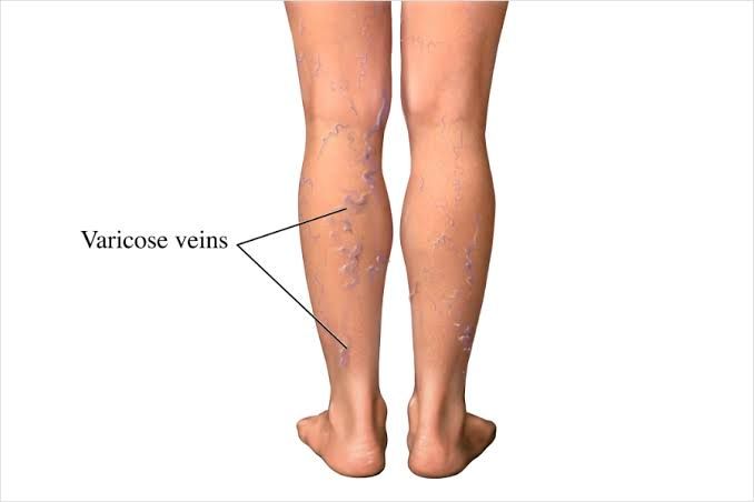
Varicose veins are enlarged, twisted, and often noticeable veins, most commonly occurring in the legs. They are a manifestation of chronic venous insufficiency.
Pathogenesis of Varicose Veins
The formation of varicose veins is primarily a consequence of compromised venous blood flow, leading to increased pressure within the veins (venous hypertension). The key mechanisms involve:
- Valve Incompetence: Veins, particularly in the limbs, contain one-way valves that prevent the backflow (reflux) of blood and help propel it towards the heart against gravity. The primary event in the development of most varicose veins is the failure or incompetence of these valves. This can be due to:
- Primary Valve Dysfunction: Intrinsic weakness of the valve structure or vein wall, often with a genetic predisposition.
- Secondary Valve Dysfunction: Damage caused by previous deep vein thrombosis (DVT), inflammation (phlebitis), or conditions that increase pressure within the abdomen (like pregnancy, obesity, or tumors), which impede venous return and stretch the vein walls, pulling the valve leaflets apart.
- Venous Wall Weakness: The walls of veins can become weakened or lose elasticity over time, causing them to dilate. This dilation further exacerbates valve incompetence as the valve leaflets can no longer meet properly.
- Increased Intraluminal Pressure: Incompetent valves lead to reflux, meaning blood flows backward down the vein segment, pooling and increasing pressure distal to the faulty valve. This elevated hydrostatic pressure stretches the vein walls, causing further dilation and tortuosity characteristic of varicose veins. In standing individuals, gravity significantly contributes to this pressure build-up in the lower limbs.
- Contributing Factors: Various factors can predispose an individual to developing varicose veins or worsen existing ones:
- Genetics: Family history is a significant risk factor.
- Age: Veins and valves can weaken over time.
- Sex: Women are more commonly affected, linked to hormonal changes (pregnancy, menopause, hormone therapy) which can relax vein walls.
- Pregnancy: Hormonal effects combined with increased blood volume and pressure on pelvic veins from the growing uterus contribute.
- Obesity: Increased abdominal pressure and overall strain on the circulatory system.
- Prolonged Standing or Sitting: Lack of muscle pump action (calf muscles contracting to help push blood up) leads to blood pooling.
The process is progressive: valve incompetence leads to reflux and dilation, which in turn worsens valve incompetence, creating a vicious cycle that results in the characteristic appearance of varicose veins.
Different Sites Where Varicose Veins Can Occur
While most commonly associated with the legs, varicose veins can occur in other areas where venous pressure is elevated or venous return is compromised. The most frequent sites include:
- Lower Limbs: This is the classic location. Varicosities typically affect the superficial venous system, primarily the Great Saphenous Vein (running from the ankle up the inner thigh) and the Small Saphenous Vein (running up the back of the calf). They can also appear as smaller spider veins (telangiectasias) or reticular veins.
- Esophagus (Esophageal Varices): These occur due to portal hypertension, usually secondary to liver disease (like cirrhosis). Increased pressure in the portal vein system is transmitted to the veins in the lower esophagus, causing them to enlarge. These are particularly dangerous as they are prone to rupture and severe bleeding.
- Rectum and Anus (Hemorrhoids): These are varicose veins of the anal and rectal venous plexuses, often caused by increased pressure from straining during defecation, pregnancy, or prolonged sitting.
- Spermatic Cord (Varicocele): Varicosities of the pampiniform plexus, a network of veins draining the testicle. Usually occurs on the left side and can affect fertility.
- Abdominal Wall (Caput Medusae): Dilated superficial veins radiating from the umbilicus, also a sign of severe portal hypertension, where collateral circulation develops to bypass the liver blockage.
While less common, varicosities can theoretically occur in other venous beds under specific conditions of localized venous obstruction or high pressure.
Sequelae (Complications) of Varicose Veins
Chronic venous hypertension resulting from varicose veins can lead to a range of complications, collectively known as chronic venous insufficiency. These include:
- Symptoms: Aching, heaviness, fatigue, throbbing, itching (venous eczema), and swelling (edema) in the affected limb, particularly after prolonged standing.
- Skin Changes: Chronic edema and inflammation lead to changes in the skin near the ankles. These can include:
- Hyperpigmentation: Brownish discoloration due to the breakdown of red blood cells that leak into the tissues.
- Venous Eczema: Itchy, dry, or weeping skin rash.
- Lipodermatosclerosis: Hardening and tightening of the skin and tissue, often giving the lower leg an inverted champagne bottle appearance.
- Atrophie Blanche: Small, white, scarred areas that are fragile.
- Phlebitis/Thrombophlebitis: Inflammation of the vein wall, often associated with the formation of a blood clot within the affected superficial vein (superficial venous thrombosis). This presents as a painful, hard, red cord along the path of the vein.
- Bleeding: Although rare, traumatized superficial varicose veins can bleed, sometimes profusely.
- Venous Ulceration: The most severe complication. Chronic high pressure impairs oxygen and nutrient delivery to the skin, making it fragile and prone to breakdown, often from minor trauma. These ulcers, known as venous stasis ulcers, typically occur in the gaiter area (above the ankle) and are slow to heal.
- Deep Vein Thrombosis (DVT): While varicose veins are superficial, severe chronic venous insufficiency can increase the risk of clots forming in the deeper veins.
Understanding Blood Vessel Tumors (Vascular Neoplasms)
Blood vessel tumors, or vascular neoplasms, are growths that arise from the cells lining blood vessels (endothelial cells) or the supporting cells around them (pericytes). They range significantly in behavior from benign growths that cause little more than cosmetic concerns to highly aggressive malignant cancers. Accurate classification and differentiation are critical for prognosis and treatment.
Criteria of Differentiation Between Benign, Borderline, and Malignant Blood Vessel Tumors
Classifying vascular tumors relies on a combination of clinical presentation, gross (macroscopic) appearance, and most importantly, microscopic (histological) features, often complemented by immunohistochemistry.
Here are the key criteria used by pathologists to grade vascular tumors:
- Clinical Behavior:
- Benign: No significant growth, slow growth, or spontaneous regression (common in infantile hemangiomas). Do not invade surrounding tissues or metastasize. Locally confined.
- Borderline: Locally aggressive tendencies, high risk of local recurrence after excision, potential for infiltrative growth. Low or very low metastatic potential.
- Malignant: Rapid growth, infiltrative and destructive of surrounding tissues. Have the capacity to metastasize (spread) to distant sites (lymph nodes, lungs, liver, etc.).
- Gross (Macroscopic) Appearance:
- Benign: Often well-circumscribed, encapsulated (though not always truly encapsulated), discrete nodule or mass. Can be soft and spongy (like cavernous hemangioma).
- Borderline: May appear more infiltrative or less clearly defined borders. Can be larger than benign counterparts.
- Malignant: Tend to be larger, poorly circumscribed with infiltrative margins, often firm consistency. May show areas of hemorrhage or necrosis (tissue death).
- Microscopic (Histological) Features: This is the most definitive level of assessment:
- Cellularity:
- Benign: Low cellularity, cells are widely separated by stroma or well-formed vascular lumens.
- Borderline: Moderate to high cellularity in areas.
- Malignant: High cellularity, densely packed cells.
- Nuclear Pleomorphism: Variation in the size, shape, and staining intensity of cell nuclei.
- Benign: Little to no nuclear pleomorphism. Nuclei are uniform.
- Borderline: Mild to moderate nuclear pleomorphism.
- Malignant: Significant nuclear pleomorphism. Nuclei are large, irregular, and hyperchromatic (darkly staining).
- Mitotic Activity: The number of cells undergoing division.
- Benign: Mitoses are rare or absent.
- Borderline: Increased mitotic activity compared to benign, but usually low to moderate numbers.
- Malignant: High mitotic activity (numerous mitoses seen per high-power field). Atypical (abnormal) mitoses may be present.
- Necrosis: Areas of dead tissue within the tumor.
- Benign: Typically absent.
- Borderline: May be present, but often focal.
- Malignant: Often present, sometimes extensive, due to rapid growth outstripping blood supply.
- Growth Pattern and Invasiveness:
- Benign: Well-formed vascular channels or spaces. May involve surrounding tissues (e.g., muscle) but do so in a non-destructive manner, respecting tissue planes.
- Borderline: Variable patterns; may show infiltrative growth into surrounding tissues, not respecting normal anatomical boundaries, but often still forming some semblance of vascular structures.
- Malignant: Destructive infiltrative growth. May show solid sheets of cells with poorly formed or no discernible vascular lumens. Cells often invade nerves or blood vessels (lympho-vascular invasion).
- Endothelial Atypia: Abnormal changes in the endothelial cells themselves.
- Benign: Endothelial cells are flat and uniform.
- Borderline: Mild to moderate endothelial cell prominence or plumpness.
- Malignant: Marked endothelial cell atypia; cells may be plump, rounded, spindle-shaped, and show significant nuclear abnormalities.
- Cellularity:
- Immunohistochemistry (IHC): Stains for specific proteins help confirm the vascular origin and can aid in classification. Common markers include CD31, CD34, and ERG, which are typically positive in endothelial cells. Other markers can help distinguish specific subtypes or rule out look-alike tumors.
A definitive diagnosis often requires microscopic examination of a biopsy specimen by a pathologist, integrating all these criteria.
Examples of Different Types of Blood Vessel Tumors
Based on the criteria above, vascular tumors are broadly categorized:
- Benign Vascular Tumors: These are common, non-cancerous growths.
- Hemangioma: The most frequent type. Can be infantile (often appearing shortly after birth and may regress spontaneously) or congenital. Subtypes include capillary hemangioma (composed of small, capillary-sized vessels, e.g., strawberry mark) and cavernous hemangioma (composed of larger, dilated vascular spaces). Can occur in skin, soft tissue, or internal organs (e.g., liver).
- Glomus Tumor: Arises from specialized glomus cells (modified smooth muscle cells associated with thermoregulation). Typically found in the extremities, especially under the fingernail, and is often intensely painful.
- Lymphangioma: Similar to hemangiomas but composed of lymphatic vessels rather than blood vessels. Can be capillary or cavernous (cystic hygroma).
- Borderline Vascular Tumors (Intermediate Malignancy): These tumors have characteristics between benign and malignant. They are locally aggressive and have a potential for recurrence but rarely metastasize. Classification within this group is complex and evolving.
- Hemangioendothelioma: A group of tumors with diverse appearances and behaviors. Examples include Epithelioid Hemangioendothelioma (can affect soft tissue, lung, liver; slow-growing but can rarely metastasize), Spindle Cell Hemangioendothelioma (often in extremities, associated with venous malformations; locally aggressive, high recurrence), and Kaposiform Hemangioendothelioma (often seen in infants/children, aggressive local growth, associated with Kasabach-Merritt phenomenon – trapping of platelets leading to severe clotting/bleeding issues).
- Malignant Vascular Tumors: These are true cancers with the potential for aggressive growth, local invasion, and distant metastasis. They are relatively rare.
- Angiosarcoma: The most common type of malignant vascular tumor. Can arise in various locations, including skin (especially in the head and neck of elderly individuals, or in limbs affected by chronic lymphedema, e.g., post-mastectomy – Stewart-Treves syndrome), soft tissue, breast, liver, spleen, or bone. Angiosarcomas are characterized by their aggressive, infiltrative growth and poor prognosis. Histologically, they display significant cellular atypia, high mitotic activity, necrosis, and destructive infiltration, often forming irregular, anastomosing (connecting) vascular channels that dissect through tissue.
Conclusion
Varicose veins and blood vessel tumors represent a spectrum of vascular pathologies requiring distinct approaches to diagnosis and management. Varicose veins, while often considered primarily a cosmetic concern, can lead to significant morbidity through chronic venous insufficiency and its complications. Blood vessel tumors, though less common, necessitate careful evaluation to differentiate benign from potentially life-threatening malignant forms. The classification of vascular tumors relies heavily on detailed histopathological assessment, guided by specific microscopic criteria and supported by immunohistochemistry.
Understanding the fundamental pathogenesis of venous disease and the critical features used in the differential diagnosis of vascular neoplasms is vital for healthcare professionals in providing appropriate patient care. Any suspected vascular abnormality warrants professional medical evaluation for accurate diagnosis and management planning.


