Thyrotoxicosis, Diffuse Hyperplasia of Thyroid, and Graves’ Disease
1. Definition and Pathogenesis of Thyrotoxicosis
Thyrotoxicosis refers to a clinical syndrome caused by elevated levels of circulating thyroid hormones (free thyroxine [FT4] and/or free triiodothyronine [FT3]), leading to a hypermetabolic state. It can result from increased production of thyroid hormones (hyperthyroidism) or the release of preformed hormones due to gland destruction (e.g., subacute thyroiditis). The most common causes include Graves’ disease, toxic multinodular goiter, and toxic adenoma. Other rarer causes include iodine-induced thyrotoxicosis and ectopic thyroid tissue.
2. Pathogenesis of Diffuse Hyperplasia of the Thyroid (Graves’ Disease)
Graves’ disease is an autoimmune disorder characterized by the production of thyroid-stimulating immunoglobulins (TSI), which mimic the action of thyroid-stimulating hormone (TSH). These autoantibodies bind to TSH receptors on thyroid follicular cells, causing continuous stimulation of hormone synthesis and secretion. This leads to diffuse hyperplasia of the thyroid gland, excessive production of T3 and T4 hormones, and clinical manifestations associated with thyrotoxicosis.
Clinical Findings in Thyrotoxicosis and Graves’ Disease:
- General Symptoms: Nervousness, anxiety, heat intolerance, increased sweating, weight loss despite increased appetite.
- Cardiovascular Symptoms: Tachycardia, atrial fibrillation (especially in older patients), systolic hypertension.
- Neurological Symptoms: Tremors, hyperactivity.
- Ophthalmopathy (specific to Graves’ disease): Proptosis (bulging eyes), periorbital edema, diplopia.
- Dermopathy: Pretibial myxedema in severe cases.
- Other Signs: Warm moist skin, muscle weakness, menstrual irregularities.
Multinodular Goiter
Definition and Pathogenesis: Multinodular goiter is characterized by an enlarged thyroid gland containing multiple nodules. It typically develops over years due to chronic stimulation by TSH in response to iodine deficiency or other factors that impair normal hormone synthesis. Over time:
- Some areas within the gland become autonomous (“hot” nodules) due to somatic mutations in genes like TSH receptor genes.
- These autonomous regions produce excess thyroid hormones independent of TSH regulation.
This condition may progress into toxic multinodular goiter if significant thyrotoxicosis develops.
Types of Solitary Thyroid Nodules
Solitary thyroid nodules are discrete lesions within the thyroid gland that differ from surrounding parenchyma. They can be classified as:
- Benign Lesions:
- Follicular adenomas
- Colloid nodules
- Cysts
- Malignant Lesions:
- Papillary carcinoma
- Follicular carcinoma
- Medullary carcinoma
- Anaplastic carcinoma
Cold vs Hot Nodules:
- A “cold” nodule does not take up radioactive iodine during scintigraphy and is often non-functional; it may indicate malignancy or benign conditions like cysts or colloid nodules.
- A “hot” nodule shows increased uptake of radioactive iodine and is usually functional or autonomous; these are less likely to be malignant.
Hypothyroidism
Clinical Findings & Pathology: Hypothyroidism results from insufficient production or action of thyroid hormones. It can be primary (thyroid dysfunction) or secondary/tertiary (pituitary/hypothalamic dysfunction).
- Symptoms: Fatigue, cold intolerance, weight gain despite poor appetite, constipation, dry skin, hair thinning/loss.
- Signs: Bradycardia, delayed reflexes (“hung-up reflexes”), puffiness around the eyes.
Cretinism: Refers to hypothyroidism in infancy/early childhood leading to impaired physical growth and mental development.
Myxedema: Severe hypothyroidism in adults characterized by generalized edema due to glycosaminoglycan accumulation in tissues.
Hashimoto’s Thyroiditis
Definition and Pathogenesis:
Hashimoto’s thyroiditis, also known as chronic lymphocytic thyroiditis, is an autoimmune disorder and the most common cause of hypothyroidism in iodine-sufficient regions. It is characterized by the immune system mistakenly attacking the thyroid gland, leading to inflammation and gradual destruction of thyroid tissue. This results in reduced production of thyroid hormones over time.
The pathogenesis involves a combination of genetic predisposition and environmental triggers. A genetic link has been established, with Hashimoto’s disease often associated with other autoimmune disorders like systemic lupus erythematosus or rheumatoid arthritis. The immune system produces high levels of autoantibodies against thyroid-specific antigens, such as thyroid peroxidase (TPO) and thyroglobulin (Tg). These antibodies lead to lymphocytic infiltration of the thyroid gland, causing follicular cell destruction and fibrosis. Over time, this process results in hypothyroidism.
Lymphocytic Thyroiditis
Definition and Pathogenesis:
Lymphocytic thyroiditis refers to a group of conditions where lymphocytes infiltrate the thyroid gland. A specific subtype is subacute lymphocytic thyroiditis, which is often seen postpartum or sporadically. This condition is considered autoimmune in origin.
In subacute lymphocytic thyroiditis, there is an initial hyperthyroid phase caused by the release of preformed T3 and T4 hormones due to follicular cell damage. This phase is followed by a hypothyroid phase as hormone stores are depleted. Antimicrosomal antibodies (anti-TPO) are present in 50-80% of patients with this condition. The exact mechanism involves immune-mediated damage but without significant pain or tenderness typically seen in other forms like subacute granulomatous thyroiditis.
Subacute Thyroiditis
Definition and Pathogenesis:
Subacute thyroiditis (also called de Quervain’s or granulomatous thyroiditis) is a self-limited inflammatory condition that typically follows a viral upper respiratory infection. It is characterized by painful swelling of the thyroid gland.
The pathogenesis involves viral infection triggering an inflammatory response within the thyroid gland. Commonly implicated viruses include mumps virus, coxsackievirus, Epstein-Barr virus, influenza virus, and adenovirus. The inflammation leads to disruption of follicular cells and leakage of stored T3 and T4 into circulation, resulting in transient hyperthyroidism during the early phase. As inflammation subsides, there may be a temporary hypothyroid phase before recovery occurs.
Histologically, subacute granulomatous thyroiditis shows multinucleated giant cells surrounding colloid material from disrupted follicles.
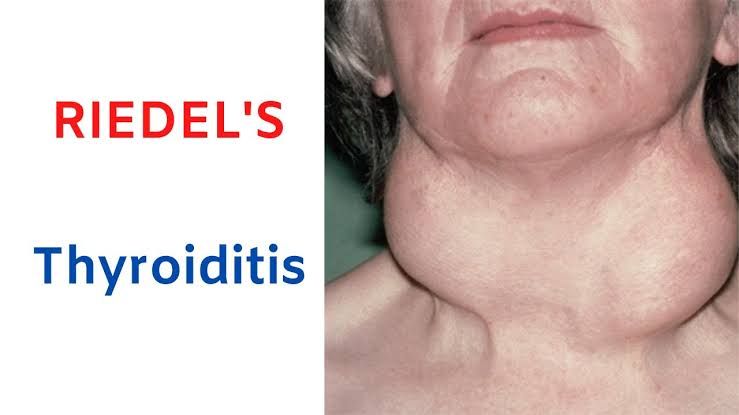
Riedel’s Thyroiditis
Definition and Pathogenesis:
Riedel’s thyroiditis is a rare form of chronic inflammatory disease characterized by extensive fibrosis that replaces normal thyroid tissue. It can extend beyond the gland into surrounding neck structures.
The exact cause remains unclear but appears to involve an autoimmune component linked to systemic fibrosclerotic syndromes such as retroperitoneal fibrosis or sclerosing cholangitis. The fibrosis leads to a “woody” hard mass in the neck that may compress adjacent structures like the trachea or esophagus. Unlike other forms of thyroiditis, Riedel’s does not primarily involve lymphocytic infiltration but rather dense fibrotic tissue formation.


