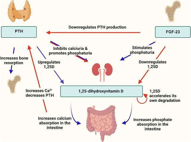
Absorption, Metabolism, and Excretion of Calcium and Phosphate
Calcium Absorption, Metabolism, and Excretion:
- Absorption: Calcium is absorbed primarily in the small intestine. The process occurs through two mechanisms:
- Active Transport (Transcellular): This is a vitamin D-dependent process that occurs mainly in the duodenum. Active calcium absorption involves calcium channels on the apical membrane of enterocytes, intracellular binding to calbindin-D proteins, and extrusion into the bloodstream via calcium ATPases or sodium-calcium exchangers.
- Passive Diffusion (Paracellular): This occurs throughout the small intestine and is independent of vitamin D. It depends on the concentration gradient between intestinal lumen and blood.
- Metabolism: Once absorbed, calcium enters the bloodstream where it exists in three forms:
- Free ionized calcium (~50%, biologically active).
- Bound to albumin (~40%).
- Complexed with anions like phosphate (~10%).
- Excretion: Calcium is excreted primarily through urine and feces. In the kidneys, about 99% of filtered calcium is reabsorbed along the nephron, particularly in the proximal tubule and loop of Henle. Parathyroid hormone (PTH) enhances renal reabsorption of calcium.
Phosphate Absorption, Metabolism, and Excretion:
- Absorption: Phosphate absorption occurs predominantly in the small intestine via sodium-phosphate cotransporters (NaPi-IIb). Approximately 30% of phosphate absorption is regulated by vitamin D.
- Metabolism: About 85% of total body phosphate is stored in bone as hydroxyapatite crystals. The remaining phosphate exists in extracellular fluids (1%) or within cells (14%). In plasma, phosphate exists as monovalent (H₂PO₄⁻) or divalent ions (HPO₄²⁻), depending on pH.
- Excretion: Phosphate homeostasis is maintained by renal excretion. Around 80–90% of filtered phosphate is reabsorbed in the proximal tubule via NaPi-IIa/IIc transporters. PTH reduces renal reabsorption by promoting internalization and degradation of these transporters.
Role of Vitamin D in Calcium and Phosphate Absorption
Vitamin D plays a critical role in maintaining calcium and phosphate homeostasis:
- Calcium Absorption:
- Vitamin D increases intestinal absorption of calcium by upregulating calbindin-D proteins in enterocytes.
- Calbindin facilitates transcellular transport of calcium from the intestinal lumen to the bloodstream.
- Without sufficient vitamin D, active calcium absorption decreases significantly.
- Phosphate Absorption:
- Vitamin D enhances intestinal phosphate absorption by increasing expression of sodium-phosphate cotransporters (NaPi-IIb) on enterocyte membranes.
- This ensures adequate availability of phosphate for bone mineralization and other cellular functions.
- Regulation Mechanism:
- Vitamin D synthesis begins with UV-induced conversion of 7-dehydrocholesterol to cholecalciferol (vitamin D₃) in skin.
- In the liver, cholecalciferol is hydroxylated to form 25-hydroxyvitamin D₃ [25(OH)D₃], which is further converted to its active form—1,25-dihydroxyvitamin D₃ [1,25(OH)₂D₃]—by renal enzyme CYP27B1 under PTH stimulation.
Effect of Calcium Ion Concentration on Regulation of Active Vitamin D Levels
The regulation of active vitamin D levels ([1,25(OH)₂D₃]) depends heavily on serum calcium concentrations:
- When serum calcium levels are low:
- Hypocalcemia stimulates PTH secretion from parathyroid glands.
- PTH upregulates CYP27B1 activity in renal proximal tubules, increasing conversion of inactive vitamin D [25(OH)D₃] into its active form [1,25(OH)₂D₃].
- Active vitamin D enhances intestinal absorption of both calcium and phosphate to restore normal serum levels.
- When serum calcium levels are high:
- Hypercalcemia suppresses PTH secretion via negative feedback mechanisms involving activation of calcium-sensing receptors (CaSRs) on parathyroid cells.
- Reduced PTH levels decrease CYP27B1 activity while increasing CYP24A1 activity (responsible for degrading active vitamin D into inactive metabolites like calcitroic acid).
This dynamic regulation ensures that excessive production or depletion of active vitamin D does not occur under normal physiological conditions.
Major Physiological Effects of Parathyroid Hormone (PTH)
PTH has several key physiological effects aimed at maintaining serum calcium homeostasis:
- Bone Resorption:
- PTH binds to receptors on osteoblasts to stimulate expression of RANKL protein.
- RANKL promotes differentiation and activation of osteoclasts, leading to bone resorption and release of stored calcium/phosphate into circulation.
- Renal Effects:
- Enhances reabsorption of calcium in distal convoluted tubules while promoting excretion of phosphate by reducing NaPi-IIa/IIc transporter activity.
- Stimulates production of active vitamin D [1,25(OH)₂D₃] by upregulating renal CYP27B1 enzyme.
- Intestinal Effects:
- Indirectly increases intestinal absorption of both calcium and phosphate through stimulation of active vitamin D synthesis.
- Inhibition Feedback Loop:
- Elevated levels of circulating PTH are downregulated by increased serum ionized calcium concentrations via CaSR-mediated feedback inhibition.
Regulation of Parathyroid Hormone Secretion
The secretion rate for PTH depends primarily on extracellular ionized calcium concentrations ([Ca²⁺]e):
- Low [Ca²⁺]e (< normal range):
- Hypocalcemia reduces activation at CaSRs located on parathyroid gland chief cells.
- This disinhibition leads to increased synthesis/release rates for mature PTH stored within secretory granules.
- High [Ca²⁺]e (> normal range):
- Hypercalcemia activates CaSRs strongly enough that intracellular signaling pathways suppress further release/synthesis activities related toward new/matured-PHT molecules respectively!
Other factors influencing secretion include: Serum Pi-level changes indirectly modulate abundance-mRNA stability-transcription-rates!


