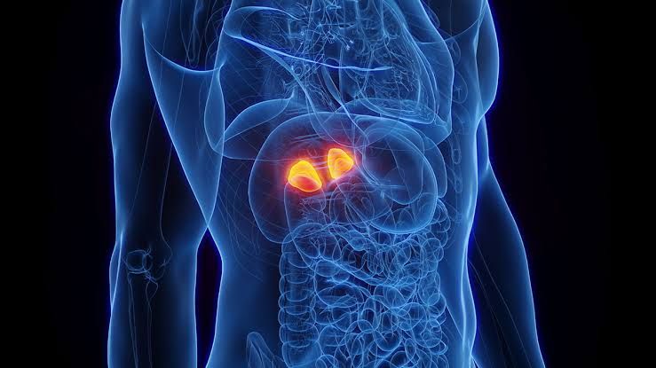
Pathological Features of Benign and Malignant Tumors of the Adrenal Gland
Benign Tumors (Adrenal Adenomas):
- Gross Appearance:
- Adrenal adenomas are typically well-circumscribed, encapsulated, and solitary.
- They have a homogeneous yellow cut surface due to their lipid-rich content.
- Necrosis and hemorrhage are rare in benign adenomas.
- Microscopic Features:
- Cells resemble normal adrenal cortical cells but may show mild atypia.
- The tumor is composed of uniform cells with clear or eosinophilic cytoplasm arranged in nests or cords.
- Mitotic activity is minimal, and there is no evidence of invasion into surrounding tissues or vasculature.
- Functional Characteristics:
- Most adrenal adenomas are nonfunctional; however, about 15% are functional and secrete hormones such as cortisol (causing Cushing’s syndrome) or aldosterone (causing primary aldosteronism).
Malignant Tumors (Adrenocortical Carcinomas):
- Gross Appearance:
- Adrenocortical carcinomas (ACCs) are larger than benign adenomas, often exceeding 5 cm in size.
- They exhibit irregular borders, areas of necrosis, hemorrhage, and cystic degeneration.
- The tumors are not encapsulated and may invade adjacent structures.
- Microscopic Features:
- Cells show marked pleomorphism with hyperchromatic nuclei and increased mitotic activity.
- Evidence of vascular invasion, capsular invasion, and necrosis is common.
- Atypical mitoses may be present.
- Functional Characteristics:
- Many ACCs are functional and produce excess steroid hormones such as cortisol, aldosterone, or sex steroids.
- Hormonal overproduction can lead to clinical syndromes like virilization or feminization.
- Prognostic Indicators:
- Poor prognosis is associated with large tumor size (>10 cm), high-grade histology (e.g., Ki-67 proliferation index >20%), and evidence of metastasis at diagnosis.
Causes of Addison’s Disease and Their Pathological Features
Causes of Addison’s Disease:
- Autoimmune Destruction:
- The most common cause in developed countries is autoimmune adrenalitis where the immune system targets the adrenal cortex.
- Associated with other autoimmune conditions like type 1 diabetes or thyroiditis (part of autoimmune polyglandular syndromes).
- Infections:
- Tuberculosis historically was a leading cause worldwide; granulomatous inflammation destroys the adrenal glands.
- Other infections include fungal infections (e.g., histoplasmosis) or HIV-related opportunistic infections.
- Metastases:
- Secondary involvement from cancers such as lung cancer, breast cancer, or melanoma can destroy adrenal tissue.
- Adrenal Hemorrhage:
- Seen in conditions like Waterhouse-Friderichsen syndrome caused by meningococcemia or anticoagulant therapy-induced bleeding into the glands.
- Genetic Disorders:
- Congenital adrenal hyperplasia due to enzyme deficiencies can lead to inadequate hormone production over time.
Pathological Features of Addison’s Disease:
- Autoimmune Adrenalitis:
- Lymphocytic infiltration replaces normal cortical tissue.
- Atrophy of all three layers of the adrenal cortex occurs while the medulla remains intact.
- Tuberculous Adrenalitis:
- Caseating granulomas replace normal glandular tissue.
- Metastatic Involvement:
- Tumor cells infiltrate the gland causing destruction.
- Hemorrhagic Necrosis:
- Extensive hemorrhage leads to gland destruction; seen in acute cases like Waterhouse-Friderichsen syndrome.
Classification of Multiple Endocrine Neoplasia (MEN)
Multiple endocrine neoplasia (MEN) is a group of rare, inherited disorders that involve the development of tumors or excessive growth in multiple endocrine glands. These conditions are classified into four main types based on the genetic mutations involved, the glands affected, and the clinical manifestations. Below is a detailed classification:
1. MEN Type 1 (MEN1)
- Gene Involved: Mutations in the MEN1 gene, which encodes for the tumor suppressor protein menin.
- Inheritance Pattern: Autosomal dominant.
- Primary Affected Glands:
- Parathyroid Glands: Over 90% of individuals develop hyperparathyroidism due to parathyroid tumors, leading to elevated calcium levels (hypercalcemia), kidney stones, and bone thinning.
- Pancreas: Tumors in pancreatic islet cells often produce hormones like gastrin (causing Zollinger-Ellison syndrome with peptic ulcers) or insulin (causing hypoglycemia).
- Pituitary Gland: Tumors may secrete prolactin (prolactinomas), growth hormone (causing acromegaly), or corticotropin (leading to Cushing syndrome).
- Other Possible Tumors:
- Adrenal gland tumors.
- Non-endocrine tumors such as lipomas or angiofibromas.
2. MEN Type 2 (MEN2)
This type is further divided into three subtypes based on specific clinical features and risks.
a. MEN Type 2A
- Gene Involved: Mutations in the RET proto-oncogene.
- Primary Features:
- Medullary thyroid carcinoma (MTC) occurs in all cases and arises from C cells producing calcitonin.
- Pheochromocytomas occur in about half of cases, causing excessive adrenaline production and symptoms like high blood pressure and palpitations.
- Hyperparathyroidism due to parathyroid hyperplasia or adenomas.
b. MEN Type 2B
- Gene Involved: Specific mutations in the RET gene.
- Distinctive Features:
- Medullary thyroid carcinoma (MTC) develops early and aggressively.
- Pheochromocytomas are common.
- No hyperparathyroidism.
- Additional physical features include mucosal neuromas (benign nerve tissue growths) and a marfanoid body habitus (tall stature with long limbs).
c. Familial Medullary Thyroid Carcinoma (FMTC)
- A subtype of MEN2 where medullary thyroid carcinoma (MTC) is the only manifestation without other associated endocrine abnormalities.
3. MEN Type 4
- Gene Involved: Mutations in the CDKN1B gene, which encodes for p27, another tumor suppressor protein.
- Inheritance Pattern: Autosomal dominant.
- Clinical Features:
- Similar to MEN1 but caused by a different genetic mutation.
- Commonly involves hyperparathyroidism followed by pituitary tumors and other endocrine or non-endocrine tumors.


