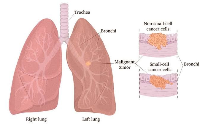
Etiology of Lung Cancer
Lung cancer arises primarily due to genetic mutations and environmental exposures. The most significant risk factor is cigarette smoking, which accounts for approximately 85-90% of lung cancer cases. Tobacco smoke contains numerous carcinogens, such as polycyclic aromatic hydrocarbons (PAHs) and nitrosamines, which induce DNA damage in respiratory epithelial cells. Other risk factors include exposure to radon gas, asbestos, air pollution (e.g., fine particulate matter), occupational hazards (e.g., arsenic or chromium), and secondhand smoke. Genetic predispositions also play a role, particularly in individuals who develop lung cancer despite never smoking.
In non-smokers, lung cancer is often associated with specific genetic mutations such as EGFR (epidermal growth factor receptor) mutations or ALK (anaplastic lymphoma kinase) rearrangements. These molecular alterations are more common in adenocarcinoma, the predominant subtype of lung cancer among never-smokers.
Small Cell Carcinoma vs. Non-Small Cell Carcinoma
Small Cell Lung Carcinoma (SCLC):
- Clinical Features: SCLC accounts for approximately 10-15% of all lung cancers and is strongly linked to smoking. It is characterized by rapid growth, early metastasis, and a strong association with paraneoplastic syndromes such as the syndrome of inappropriate antidiuretic hormone secretion (SIADH) or ectopic ACTH production.
- Pathologic Findings: Histologically, SCLC consists of small cells with scant cytoplasm, hyperchromatic nuclei, nuclear molding, and high mitotic activity. It originates from neuroendocrine cells in the bronchial epithelium.
- Prognosis: SCLC has an aggressive clinical course with a poor prognosis. Median survival without treatment is only 2-4 months; however, chemotherapy can extend survival to about 12 months in limited-stage disease.
Non-Small Cell Lung Carcinoma (NSCLC):
NSCLC constitutes about 85% of all lung cancers and includes three main subtypes:
- Adenocarcinoma:
- Most common subtype overall and especially prevalent among non-smokers.
- Arises from glandular epithelial cells; often located peripherally in the lungs.
- Pathology shows gland formation or mucin production.
- Prognosis varies depending on stage but tends to be better than other NSCLCs if detected early.
- Squamous Cell Carcinoma:
- Strongly associated with smoking; typically arises centrally near large airways.
- Histology reveals keratin pearls and intercellular bridges.
- Often presents with symptoms like hemoptysis due to central airway involvement.
- Prognosis depends on stage but is generally worse than adenocarcinoma.
- Large Cell Carcinoma:
- Poorly differentiated tumor lacking features of adenocarcinoma or squamous cell carcinoma.
- Can occur anywhere in the lungs; histology shows large pleomorphic cells without glandular or squamous differentiation.
- Prognosis is poor due to its aggressive nature.
Bronchial Carcinoid Tumors
Bronchial carcinoid tumors are rare neuroendocrine tumors that account for less than 5% of all primary lung cancers. They are classified into two types:
- Typical Carcinoids: Low-grade tumors with minimal mitotic activity and no necrosis.
- Atypical Carcinoids: Intermediate-grade tumors with higher mitotic rates and focal necrosis.
These tumors commonly arise centrally within the bronchi and may cause symptoms such as cough, hemoptysis, wheezing, or recurrent infections due to airway obstruction. Unlike SCLC, bronchial carcinoids rarely metastasize but can secrete hormones leading to paraneoplastic syndromes like carcinoid syndrome (flushing, diarrhea). Surgical resection offers excellent long-term outcomes for typical carcinoids.
Paraneoplastic Syndromes Associated with Lung Cancer
Paraneoplastic syndromes are systemic effects caused by substances secreted by tumors or immune cross-reactivity between tumor antigens and normal tissues:
- Endocrine Syndromes:
- SIADH: Commonly seen in SCLC; leads to hyponatremia due to inappropriate ADH secretion.
- Ectopic Cushing’s Syndrome: Caused by ACTH secretion from SCLC.
- Hypercalcemia: Frequently associated with squamous cell carcinoma due to parathyroid hormone-related protein (PTHrP) secretion.
- Neurologic Syndromes:
- Lambert-Eaton Myasthenic Syndrome: Autoimmune disorder causing muscle weakness; associated with SCLC.
- Dermatologic Syndromes:
- Acanthosis Nigricans: Rarely seen but can occur as a paraneoplastic phenomenon.
- Hematologic Syndromes:
- Trousseau’s Syndrome: Hypercoagulability leading to venous thrombosis; often linked to adenocarcinomas.
- Others: Hypertrophic osteoarthropathy causing clubbing of fingers may be seen in NSCLC.
Treatment involves addressing both the underlying malignancy and managing specific symptoms caused by these syndromes.
Other Tumors in the Lung
Primary Tumors:
- Besides NSCLC and SCLC, other rare primary lung tumors include sarcomas (e.g., pulmonary leiomyosarcoma), lymphomas involving the lungs primarily or secondarily, and benign lesions like hamartomas.
Metastatic Tumors:
The lungs are a common site for metastatic spread because they receive blood from both systemic circulation via pulmonary arteries/veins:
- The most frequent metastatic tumors originate from breast cancer, colorectal cancer, renal cell carcinoma, melanoma, sarcomas, and head/neck cancers.
Metastases typically present as multiple nodules scattered throughout both lungs on imaging studies.
Diagnostic Techniques Used for Respiratory Disease
The diagnostic techniques used for respiratory diseases are as follows:
- Physical Examination:
- Auscultation using a stethoscope to detect abnormal breath sounds such as wheezing, crackles, or diminished breath sounds.
- Percussion of the chest to assess fluid accumulation or air trapping.
- Imaging Studies:
- Chest X-ray: Commonly used to identify abnormalities like pneumonia, pleural effusion, pneumothorax, or tumors.
- Computed Tomography (CT) Scan: Provides detailed images of the lungs and is useful for detecting small nodules, masses, or interstitial lung disease.
- Magnetic Resonance Imaging (MRI): Occasionally used for soft tissue evaluation in cases of suspected tumors.
- Ultrasound: Useful for detecting pleural effusions and guiding thoracentesis.
- Pulmonary Function Tests (PFTs):
- Measures lung capacity, airflow, and gas exchange efficiency.
- Includes spirometry (to measure forced expiratory volume and vital capacity) and diffusion capacity tests.
- Bronchoscopy:
- A flexible or rigid scope is inserted into the airways to visualize abnormalities directly.
- Allows biopsy collection for histopathological analysis.
- Thoracentesis:
- A needle is inserted into the pleural space to remove fluid for diagnostic purposes in cases of pleural effusion.
- Biopsy:
- Tissue samples from the lung, pleura, or lymph nodes are obtained via bronchoscopy, CT-guided needle biopsy, or surgical methods.
- Blood Tests:
- Microbiological Studies:
- Sputum culture and sensitivity testing identify infectious organisms like bacteria or fungi.
- Acid-fast bacilli staining and culture are performed for tuberculosis diagnosis.
- Pleural Fluid Analysis:
- Examines fluid obtained through thoracentesis to determine if it is transudative (e.g., due to heart failure) or exudative (e.g., due to infection or malignancy).
- Molecular Testing:
- Genetic testing for mutations such as EGFR in lung cancer patients helps guide targeted therapy decisions.
- Pulse Oximetry and Capnography:
- Non-invasive methods to monitor oxygen saturation and carbon dioxide levels in real-time.
Pleural Effusions, Pneumothorax, and Pleural Tumors
Pleural Effusions:
- Accumulation of excess fluid in the pleural space.
- Causes include congestive heart failure (transudative), infections like pneumonia (exudative), malignancies, pulmonary embolism, or autoimmune diseases.
- Symptoms include dyspnea, chest pain, and reduced breath sounds on the affected side.
- Diagnosis involves imaging studies like chest X-rays or ultrasound followed by thoracentesis for fluid analysis.
Pneumothorax:
- Air accumulation in the pleural space leading to partial or complete lung collapse.
- Types:
- Spontaneous pneumothorax (primary without underlying disease; secondary with conditions like COPD).
- Traumatic pneumothorax caused by injury.
- Tension pneumothorax—a medical emergency where trapped air compresses mediastinal structures.
- Symptoms include sudden chest pain and dyspnea; diagnosis is confirmed via chest X-ray.
Pleural Tumors:
- Include benign lesions like fibrous tumors of the pleura and malignant mesothelioma associated with asbestos exposure.
- Symptoms may involve chest pain, dyspnea, weight loss, and recurrent pleural effusions.
Nasal Polyp, Papilloma, and Carcinoma
Nasal Polyp:
- Benign growths arising from inflamed mucosa within nasal passages or sinuses due to chronic inflammation from allergies or infections.
- Symptoms include nasal obstruction, anosmia (loss of smell), rhinorrhea (runny nose), and sinusitis-like symptoms.
Nasal Papilloma:
- Benign epithelial tumors caused by human papillomavirus (HPV).
- Inverted papillomas are locally aggressive with a risk of malignant transformation into squamous cell carcinoma.
Nasal Carcinoma:
- Malignant tumors arising from nasal cavity epithelium; squamous cell carcinoma is most common.
- Risk factors include smoking exposure to industrial chemicals like nickel dust.
Etiology and Pathology of Nasopharyngeal Carcinoma
Etiology: Nasopharyngeal carcinoma has a strong association with Epstein-Barr Virus (EBV) infection along with genetic predisposition and environmental factors such as consumption of salted fish containing nitrosamines.
Pathology: This carcinoma arises from nasopharyngeal epithelium often presenting as undifferentiated nonkeratinizing carcinoma infiltrating local tissues early due its location near lymphatic drainage pathways.
Laryngeal Polyp, Papilloma, and Carcinoma
Laryngeal Polyp:
- Benign growths on vocal cords caused by vocal strain/overuse often seen in singers (“Singer’s nodule”).
- Symptoms include hoarseness but no systemic signs like fever.
Laryngeal Papilloma:
- Caused by HPV types 6/11 leading-to benign wart-like growths on larynx mucosa potentially causing airway obstruction children adults alike.


