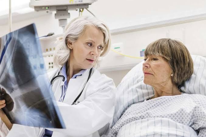
Definition of Pneumonia and Pneumonitis
Pneumonia is an inflammatory condition of the lung primarily affecting the alveoli, typically caused by infection with bacteria, viruses, fungi, or other microorganisms. It is characterized by symptoms such as fever, cough (often productive), chest pain, and difficulty breathing. The inflammation leads to alveolar filling with fluid or pus, impairing gas exchange.
Pneumonitis, on the other hand, refers to a non-infectious inflammation of lung tissue that can be caused by irritants such as chemicals, radiation therapy, allergens, or autoimmune conditions. Unlike pneumonia, pneumonitis does not necessarily involve microbial infection but instead results from exposure to external agents or immune-mediated processes.
Classification of Pneumonias According to Etiology and Morphological Patterns
Etiological Classification
- Bacterial Pneumonia: Caused by bacterial pathogens such as Streptococcus pneumoniae, Haemophilus influenzae, Klebsiella pneumoniae, and Staphylococcus aureus.
- Viral Pneumonia: Caused by respiratory viruses like influenza virus, respiratory syncytial virus (RSV), adenovirus, and coronaviruses (e.g., SARS-CoV-2).
- Fungal Pneumonia: Caused by fungi such as Aspergillus, Histoplasma capsulatum, Cryptococcus neoformans, and Coccidioides immitis.
- Aspiration Pneumonia: Results from inhalation of oropharyngeal or gastric contents containing bacteria into the lungs.
- Atypical Pneumonia: Often caused by organisms like Mycoplasma pneumoniae, Chlamydia pneumoniae, and Legionella pneumophila.
- Opportunistic Pneumonia: Occurs in immunocompromised individuals due to pathogens like Pneumocystis jirovecii or cytomegalovirus (CMV).
Morphological Patterns
- Lobar Pneumonia:
- Involves consolidation of an entire lobe of the lung.
- Typically associated with infections like those caused by Streptococcus pneumoniae.
- Bronchopneumonia:
- Characterized by patchy consolidation around bronchioles.
- Commonly seen in infections caused by gram-negative bacteria or staphylococci.
- Interstitial Pneumonia:
- Involves inflammation confined to the interstitial spaces rather than alveoli.
- Frequently associated with viral infections or atypical pathogens like Mycoplasma.
Comparison Between Bacterial and Nonbacterial Pneumonias
Causes
- Bacterial Pneumonia:
- Commonly caused by Streptococcus pneumoniae (pneumococcus), which is the leading bacterial pathogen in both children and adults.
- Other causative agents include Haemophilus influenzae, Klebsiella pneumoniae, Staphylococcus aureus, Escherichia coli, and anaerobic bacteria.
- Often occurs as a primary infection or secondary to viral infections when the immune system is weakened.
- Nonbacterial Pneumonia:
- Primarily caused by viruses such as influenza virus, respiratory syncytial virus (RSV), SARS-CoV-2 (COVID-19), adenovirus, or measles virus.
- Fungal pneumonia (a subset of nonbacterial pneumonia) can result from pathogens like Histoplasma capsulatum or Aspergillus spp., but it is less common.
- Viral pneumonia often develops after exposure to respiratory droplets from infected individuals.
Symptoms
- Bacterial Pneumonia:
- Symptoms tend to have a sudden onset.
- High fever (>38.5°C), chills, productive cough with yellow/green sputum (sometimes blood-streaked), chest pain during breathing or coughing, fatigue, rapid breathing (tachypnea), and confusion in severe cases.
- Localized lung involvement is typical; one lobe may show significant inflammation on imaging.
- Nonbacterial Pneumonia:
- Symptoms are more gradual in onset compared to bacterial pneumonia.
- Fever may be lower-grade (<38°C), dry cough or mild sputum production, wheezing, fatigue, muscle aches, sore throat, nasal congestion (in viral cases).
- Diffuse lung involvement rather than localized areas of inflammation is common in viral pneumonia.
Diagnostic Methods
- Bacterial Pneumonia:
- Chest X-rays typically reveal localized opacities or consolidation in one lobe of the lung.
- Blood tests may show elevated white blood cell counts with neutrophilia.
- Sputum cultures or blood cultures can confirm the presence of specific bacterial pathogens.
- Nonbacterial Pneumonia:
- Chest X-rays show diffuse infiltrates rather than localized consolidation seen in bacterial cases.
- Viral testing through polymerase chain reaction (PCR) assays or antigen detection can identify specific viruses like influenza or SARS-CoV-2.
- Thin-section computed tomography (CT) scans may reveal centrilobular nodules in viral cases but lack sensitivity for definitive differentiation from bacterial causes.
Treatment
- Bacterial Pneumonia:
- Treated effectively with antibiotics such as beta-lactams (penicillins/cephalosporins) or macrolides depending on the pathogen’s susceptibility profile.
- Severe cases may require hospitalization for intravenous antibiotics and oxygen therapy.
- Nonbacterial Pneumonia:
- Antibiotics are ineffective against viral pathogens. Antiviral medications like oseltamivir may be used for influenza-related pneumonia if started early.
- Supportive care includes hydration, rest, fever management with antipyretics like acetaminophen, and oxygen therapy if hypoxia occurs.
Prognosis
- Bacterial Pneumonia:
- With prompt antibiotic treatment, most patients recover within days to weeks. However, complications such as pleural effusion or sepsis can occur if untreated.
- Nonbacterial Pneumonia:
- Viral pneumonias often resolve on their own within one to two weeks if uncomplicated. However, severe forms like COVID-19-associated pneumonia can lead to acute respiratory distress syndrome (ARDS) requiring intensive care support.
Events in the Resolution of the Pneumonic Process
The resolution of pneumonia involves several sequential steps:
- Edema Phase (Congestion):
- Initial response involves vascular congestion and exudation into alveoli.
- Alveoli fill with proteinaceous fluid but contain few inflammatory cells.
- Red Hepatization Phase:
- Marked infiltration of neutrophils into alveolar spaces along with fibrin deposition.
- The lung appears red and firm due to capillary congestion.
- Gray Hepatization Phase:
- Neutrophils are replaced by macrophages that begin phagocytosing cellular debris.
- Fibrin persists while capillary congestion subsides.
- Resolution Phase:
- Macrophages clear remaining debris through phagocytosis.
- Normal lung architecture is restored if no complications occur.
Possible Complications of Pneumonia
- Respiratory failure due to impaired gas exchange.
- Pleural effusion (fluid accumulation in pleural space).
- Empyema (infected pleural effusion).
- Lung abscess formation due to localized necrosis.
- Sepsis resulting from systemic spread of infection.
- Chronic complications such as bronchiectasis.
Causes, Morphology, and Outcome of Lung Abscess
Causes
- Aspiration of oral secretions (common in alcoholics or patients with altered consciousness).
- Post-obstructive infections secondary to bronchial obstruction (tumors/foreign bodies).
- Hematogenous spread from septic emboli (e.g., infective endocarditis).
- Direct extension from adjacent infections (subphrenic abscess).
Morphology
- Gross Appearance:
- Cavitary lesion > 2 cm filled with pus or necrotic debris within lung parenchyma.
- Surrounded by inflamed tissue; may show air-fluid levels on imaging if connected to bronchi.
- Microscopic Features:
- Central necrotic area containing neutrophils and bacteria surrounded by granulation tissue.
Outcome
- Favorable outcomes include resolution after antibiotic therapy with scar formation at the site of abscesses.
- Adverse outcomes include chronic abscess formation, rupture into pleura causing empyema/pyopneumothorax, sepsis, or death if untreated.


