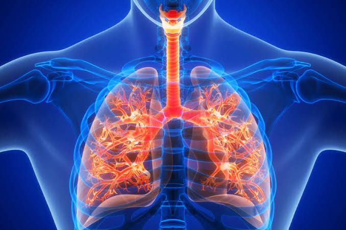
Function of the Dorsal and Ventral Groups of Respiratory Neurons, the Pneumotaxic Center, and the Apneustic Center in the Brain Stem
The respiratory centers in the brainstem are critical for controlling breathing. These centers include the dorsal respiratory group (DRG), ventral respiratory group (VRG), pneumotaxic center, and apneustic center.
1. Dorsal Respiratory Group (DRG)
The DRG is located in the medulla oblongata, specifically within the nucleus of the solitary tract. It primarily consists of inspiratory neurons that play a fundamental role in initiating and maintaining inspiration. The DRG receives sensory input from peripheral chemoreceptors, baroreceptors, and stretch receptors via cranial nerves such as the vagus nerve (cranial nerve X) and glossopharyngeal nerve (cranial nerve IX). This input allows it to integrate information about oxygen levels, carbon dioxide levels, blood pH, and lung stretch to regulate breathing rhythm. The DRG sends signals to motor neurons that control the diaphragm and external intercostal muscles for quiet inspiration.
2. Ventral Respiratory Group (VRG)
The VRG is also located in the medulla oblongata but lies more ventrally than the DRG. It contains both inspiratory and expiratory neurons. Unlike the DRG, which is active during quiet breathing, VRG neurons are primarily involved during forceful breathing or when metabolic demands increase (e.g., exercise). Inspiratory neurons in this group stimulate accessory muscles of inhalation, while expiratory neurons activate muscles such as internal intercostals and abdominal muscles for active exhalation. The VRG also includes specialized regions like the pre-Bötzinger complex, which is thought to act as a pacemaker for generating rhythmic breathing patterns.
3. Pneumotaxic Center
The pneumotaxic center is located in the upper part of the pons. Its primary function is to regulate the “switch-off” point of inspiration by inhibiting inspiratory signals from both medullary centers (DRG and VRG). This inhibition shortens inspiratory duration, thereby increasing respiratory rate when necessary. Strong signals from this center result in rapid shallow breaths, while weak signals allow deeper breaths with slower rates. It works antagonistically with the apneustic center to fine-tune respiratory patterns.
4. Apneustic Center
The apneustic center resides in the lower pons and promotes prolonged inspiration by providing continuous excitatory signals to inspiratory neurons in the medulla. This stimulation delays inspiratory “switch-off,” leading to deeper breaths. However, its activity is modulated by inhibitory inputs from both pulmonary stretch receptors via vagus nerves and from the pneumotaxic center.
Effects on Respiration Mediated by Vagus Nerves
The vagus nerves play a crucial role in regulating respiration through their sensory (afferent) and motor (efferent) fibers:
- Hering-Breuer Reflex: Stretch receptors in lung tissues send inhibitory signals via vagus nerves to terminate inspiration when lungs are overinflated. This prevents excessive lung expansion.
- Deflation Reflex: Sudden lung deflation triggers strong inspiratory efforts via reduced vagal activity.
- Irritant Receptor Activation: Irritant receptors stimulated by noxious substances or particles send signals through vagus nerves to induce coughing or bronchoconstriction.
- J-Receptor Stimulation: Juxtapulmonary capillary receptors respond to pulmonary congestion or edema by causing rapid shallow breathing.
- Modulation of Breathing Rhythm: Vagal afferents provide feedback to central respiratory centers about lung volume changes during each breath cycle.
- Parasympathetic Control: Efferent fibers contribute to bronchoconstriction and secretion regulation within airways.
Neural Factors That Affect Activity of Respiratory Centers
Several neural factors influence respiratory centers:
- Chemoreceptor Input:
- Central chemoreceptors detect changes in cerebrospinal fluid pH caused by CO2 levels.
- Peripheral chemoreceptors located in carotid bodies sense arterial O2 levels, CO2 levels, and pH.
- Pulmonary Stretch Receptors:
- These mechanoreceptors prevent overinflation through inhibitory feedback via vagus nerves.
- Proprioceptors:
- Found in joints, tendons, and muscles; they stimulate increased ventilation during physical activity.
- Higher Brain Centers:
- The cerebral cortex allows voluntary control over breathing (e.g., holding breath).
- The hypothalamus influences respiration during emotional states like fear or stress.
- Reflexes:
- Irritant reflexes trigger protective responses like coughing or sneezing.
- Pain stimuli can increase respiratory rate.
- Other Inputs:
- Signals from muscle spindles help adjust respiratory effort under increased load conditions.
Abnormal Patterns of Breathing
Abnormal breathing patterns can arise due to dysfunctions within central or peripheral components of respiration:
- Cheyne-Stokes Respiration:
- Characterized by cyclic waxing-and-waning tidal volumes followed by apnea periods.
- Commonly seen in heart failure or brain injuries affecting higher centers.
- Biot’s Breathing:
- Irregular clusters of rapid breaths interspersed with apnea periods.
- Often associated with damage to medullary centers due to trauma or stroke.
- Kussmaul Breathing:
- Deep labored breathing pattern seen during metabolic acidosis (e.g., diabetic ketoacidosis).
- Apneustic Breathing:
- Prolonged inspiratory phases interrupted only briefly by expirations.
- Results from damage to pontine structures like apneustic center.
- Ataxic Breathing:
- Completely irregular pattern with varying depths; indicates severe brainstem injury.
- Central Sleep Apnea:
- Periods of absent airflow due to impaired central drive for respiration.
Cough and Sneezing Reflexes
Cough Reflex:
The cough reflex is a protective mechanism designed to clear the airways of irritants, mucus, or foreign particles. It involves the following steps:
- Stimulation: Irritants such as dust, smoke, or pathogens stimulate sensory receptors in the respiratory tract, particularly in the larynx, trachea, and bronchi.
- Afferent Pathway: The sensory signals are transmitted via the vagus nerve (cranial nerve X) to the medulla oblongata in the brainstem.
- Central Processing: The medullary cough center processes these signals and initiates a response.
- Efferent Pathway: Motor signals are sent through nerves such as the phrenic nerve (to the diaphragm), intercostal nerves (to intercostal muscles), and vagus nerve (to laryngeal muscles).
- Response: A deep inhalation is followed by closure of the glottis. Then, contraction of expiratory muscles generates high intrathoracic pressure. When the glottis opens suddenly, air is expelled forcefully, clearing irritants from the airway.
Sneezing Reflex:
The sneezing reflex serves to expel irritants from the nasal passages and upper respiratory tract:
- Stimulation: Irritation of nasal mucosa by allergens, dust, or other particles activates sensory receptors.
- Afferent Pathway: Signals travel via branches of the trigeminal nerve (cranial nerve V) to the sneeze center located in the medulla oblongata.
- Central Processing: The sneeze center coordinates a response involving multiple muscle groups.
- Efferent Pathway: Motor commands are sent to respiratory muscles (via phrenic and intercostal nerves), facial muscles (via facial nerve), and laryngeal muscles (via vagus nerve).
- Response: A deep inhalation occurs first, followed by forceful exhalation through both nasal passages and mouth due to coordinated muscle contractions.
Functions of Respiratory Receptors
1. Carotid Body
- Located at the bifurcation of each common carotid artery.
- Contains peripheral chemoreceptors that primarily detect changes in arterial oxygen partial pressure (PO2), carbon dioxide partial pressure (PCO2), and pH levels.
- Functions:
- Detects hypoxemia (low PO2) and stimulates an increase in ventilation rate when PO2 drops significantly below normal levels (<60 mmHg).
- Responds to hypercapnia (elevated PCO2) and acidosis (low pH) by signaling for increased ventilation.
2. Aortic Body
- Located near the arch of the aorta.
- Similar to carotid bodies but less sensitive; also contains peripheral chemoreceptors that monitor PO2, PCO2, and pH levels.
- Functions:
- Provides additional input on blood gas levels to fine-tune ventilatory responses.
- Plays a role in detecting systemic hypoxia.
3. Ventral Surface of Medulla Oblongata
- Houses central chemoreceptors sensitive primarily to changes in pH within cerebrospinal fluid (CSF), which indirectly reflects arterial PCO2.- Functions:
- Monitors CSF pH changes caused by CO2 diffusion across the blood-brain barrier.
- Stimulates increased ventilation when CO2 levels rise or when acidosis occurs.
Effects of Arterial PO₂, PCO₂, and pH on Alveolar Ventilation
Arterial PO₂
- Low arterial oxygen partial pressure (PO2) stimulates peripheral chemoreceptors in carotid and aortic bodies when it falls below ~60 mmHg.
- This triggers an increase in alveolar ventilation to enhance oxygen uptake into blood.
Arterial PCO₂
- Elevated arterial carbon dioxide partial pressure (PCO2) is detected by both central chemoreceptors (via CSF pH changes) and peripheral chemoreceptors.
- Increased PCO2 leads to hyperventilation as a compensatory mechanism to expel excess CO2.- Conversely, low PCO2 reduces ventilatory drive.
Blood pH
- Acidosis (low blood pH due to high H+) stimulates both central and peripheral chemoreceptors to increase ventilation rate for CO2 removal, which helps restore normal pH through reduced H+.- Alkalosis (high blood pH due to low H+) suppresses ventilation temporarily until homeostasis is achieved.


