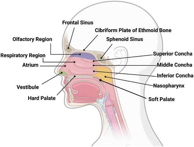
Microscopic Structure of the Upper Respiratory Passage, Including the Respiratory Mucosa
The upper respiratory passage consists of the nasal cavity, paranasal sinuses, pharynx, and the portion of the larynx above the vocal cords. These structures are lined with specialized epithelium called respiratory mucosa, which is primarily pseudostratified ciliated columnar epithelium with goblet cells.
Respiratory Mucosa
- Structure:
- The respiratory mucosa is composed of pseudostratified ciliated columnar epithelium interspersed with goblet cells. Beneath this epithelial layer lies a lamina propria containing connective tissue, blood vessels, and seromucous glands.
- Goblet cells secrete mucus that traps dust, pathogens, and other particles.
- The cilia on the epithelial cells beat rhythmically to move mucus and trapped debris toward the pharynx for swallowing or expulsion.
- Function:
- The primary function of the respiratory mucosa is to filter, warm, and humidify incoming air.
- The rich vascular network in the lamina propria helps warm inhaled air by convection.
- Mucus secreted by goblet cells and seromucous glands traps pathogens and particulate matter.
- Ciliary motion ensures that trapped debris does not accumulate in the nasal cavity or trachea but is instead directed toward the esophagus for disposal.
Correlation Between Structure and Function of Components in Nose and Trachea
Nose
- Structure:
- The nasal cavity contains three bony projections called nasal conchae (superior, middle, inferior), which increase surface area.
- The olfactory epithelium in the roof of the nasal cavity contains sensory receptors for smell.
- Paranasal sinuses are air-filled spaces lined with respiratory mucosa.
- Function:
- The increased surface area provided by conchae disrupts airflow, making it turbulent. This turbulence enhances warming, humidification, and filtration of air.
- Olfactory epithelium allows detection of airborne odorants.
- Paranasal sinuses lighten skull weight while also contributing to warming and humidifying inhaled air.
Trachea
- Structure:
- The trachea is supported by C-shaped hyaline cartilage rings that maintain its patency (open structure).
- It is lined with pseudostratified ciliated columnar epithelium containing goblet cells.
- Beneath this epithelium lies a submucosal layer containing seromucous glands.
- Function:
- Cartilage rings prevent collapse during respiration while allowing flexibility during movement or swallowing (due to posterior soft tissue).
- Mucus traps debris while cilia propel it upward toward the pharynx for clearance (the “mucociliary escalator”).
Microscopic Structure of Main Bronchi and Their Subdivisions
Main Bronchi
- Structure:
- Lined with pseudostratified ciliated columnar epithelium similar to that in the trachea but slightly thinner as bronchi branch further.
- Walls contain cartilage plates instead of complete rings as seen in the trachea. These plates provide structural support while allowing some flexibility.
- Smooth muscle fibers are present between cartilage plates.
- Function:
- Cartilage plates maintain airway patency while allowing bronchi to expand or contract slightly during breathing.
- Smooth muscle regulates bronchial diameter through bronchoconstriction or bronchodilation.
Subdivisions: Secondary (Lobar) Bronchi → Tertiary (Segmental) Bronchi → Bronchioles
- As bronchi branch into smaller subdivisions:
- Epithelium transitions from pseudostratified columnar to simple columnar or cuboidal in terminal bronchioles.
- Goblet cells decrease in number; club cells appear in terminal bronchioles to secrete surfactant-like substances.
- Functionally:
- Smaller bronchioles lack cartilage but have smooth muscle that controls airflow resistance via constriction/dilation.
Microscopic Structure of Lung Parenchyma
Alveoli
- Structure:
- Alveoli are small sac-like structures composed primarily of two cell types:
- Type I alveolar cells: Thin squamous epithelial cells forming most (97%) of alveolar surface area. They facilitate gas exchange due to their minimal thickness (~25 nm).
- Type II alveolar cells: Cuboidal epithelial cells interspersed among Type I cells. They produce pulmonary surfactant—a phospholipid-protein mixture that reduces surface tension within alveoli to prevent collapse during exhalation.
- Alveolar macrophages patrol alveolar surfaces to remove debris/pathogens.
- Alveoli are small sac-like structures composed primarily of two cell types:
Alveoli are surrounded by an extensive capillary network separated from alveolar walls by a thin basement membrane (~0.5 µm thick). Together they form a “respiratory membrane.”
- Function:
- Gas exchange occurs across this thin respiratory membrane via diffusion:
- Oxygen diffuses from alveolar air into capillary blood due to partial pressure gradients.
- Carbon dioxide diffuses from blood into alveoli for exhalation.
- Gas exchange occurs across this thin respiratory membrane via diffusion:
Alveolar Ducts/Sacs
- Alveolar ducts connect respiratory bronchioles to clusters of alveoli called alveolar sacs. These ducts/sacs maximize surface area for gas exchange.
Correlation Between Lung Parenchyma Structure and Gas Exchange Function
- Thin Respiratory Membrane:
- Facilitates rapid diffusion due to minimal barrier thickness (~0.5 µm).
- Large Surface Area:
- Each lung contains ~300 million alveoli providing ~70 m² total surface area—critical for efficient oxygen-carbon dioxide exchange.
- Elasticity:
- Elastic fibers surrounding each alveolus allow expansion during inhalation and recoil during exhalation.
- Surfactant Production by Type II Cells:
- Reduces surface tension within alveoli preventing collapse at low lung volumes.
- Capillary-Alveolus Proximity:
- Close association ensures efficient oxygen uptake into blood and carbon dioxide removal.


