Mechanics of Pulmonary Ventilation
Pulmonary ventilation, or breathing, is the process of moving air into and out of the lungs to facilitate gas exchange. It involves two primary phases: inspiration (inhalation) and expiration (exhalation). The mechanics of pulmonary ventilation are driven by pressure gradients created by changes in lung volume, which follow Boyle’s Law: pressure is inversely proportional to volume.
- Inspiration:
- During inspiration, the diaphragm contracts and moves downward, while the external intercostal muscles contract to lift the rib cage upward and outward.
- These actions increase the thoracic cavity’s volume, causing a decrease in intrapulmonary (alveolar) pressure below atmospheric pressure.
- Air flows into the lungs because air moves from areas of higher pressure (atmosphere) to lower pressure (alveoli).
- Expiration:
- Expiration is typically passive during normal breathing. The diaphragm and intercostal muscles relax, reducing thoracic cavity volume.
- This increases alveolar pressure above atmospheric pressure, forcing air out of the lungs.
- In forced expiration (e.g., during exercise), accessory muscles such as abdominal muscles contract to further reduce thoracic volume.
The movement of air depends on three pressures: pleural pressure, alveolar pressure, and transpulmonary pressure.
Definitions of Key Pressures
- Pleural Pressure:
- Pleural pressure is the pressure within the pleural cavity (the space between the visceral and parietal pleurae).
- It is always negative relative to atmospheric pressure due to opposing forces: lung elastic recoil pulling inward and chest wall elasticity pulling outward.
- Normal pleural pressure at rest is approximately -4 mm Hg.
- Alveolar Pressure:
- Alveolar pressure refers to the air pressure within the alveoli.
- It fluctuates during breathing: it decreases below atmospheric pressure during inspiration (to allow airflow into the lungs) and increases above atmospheric pressure during expiration (to expel air).
- Transpulmonary Pressure:
- Transpulmonary pressure is defined as the difference between alveolar pressure (Palv) and pleural pressure (Ppl). Ptp = Palv − Ppl. This gradient determines lung expansion; a higher transpulmonary pressure corresponds to greater lung inflation.
Changes in Lung Volumes and Pressures During Normal Breathing
- Inspiration:
- Lung Volume: Increases as thoracic cavity expands.
- Pleural Pressure: Becomes more negative (e.g., from −4 mm Hg at rest to about −6 mm Hg).
- Alveolar Pressure: Drops slightly below atmospheric (−1 mm Hg), creating a gradient for airflow into the lungs.
- Transpulmonary Pressure: Increases due to more negative pleural pressures, promoting lung expansion.
- Expiration:
- Lung Volume: Decreases as thoracic cavity recoils.
- Pleural Pressure: Returns toward resting levels (−4 mm Hg).
- Alveolar Pressure: Rises above atmospheric (+1 mm Hg), driving air out of the lungs.
- Transpulmonary Pressure: Decreases as pleural pressures become less negative.
Compliance of the Lungs
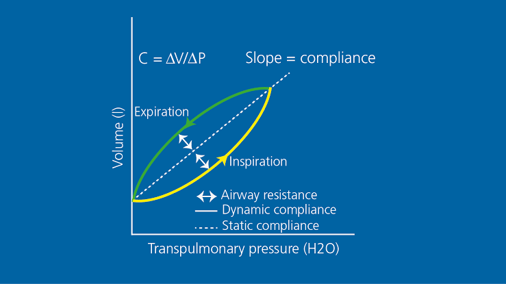
Lung compliance refers to how easily the lungs can expand in response to changes in transpulmonary pressure. It is mathematically expressed as:
C = ΔV ÷ ΔP
Where C is compliance, ΔV is change in lung volume, and ΔP is change in transpulmonary pressure.
- High compliance indicates that less effort is needed for lung expansion (e.g., emphysema).
- Low compliance means greater effort is required for expansion due to stiff or non-elastic tissue (e.g., pulmonary fibrosis).
Compliance Diagram of Lungs in a Normal Person
A compliance diagram plots lung volume against transpulmonary pressures during inspiration and expiration:
- The curve shows hysteresis—lung compliance differs between inspiration and expiration due to surface tension effects within alveoli.
- At low volumes, compliance is high because alveoli are easier to inflate when partially collapsed.
- At high volumes, compliance decreases because elastic fibers resist further stretching.
The slope of this curve represents dynamic or static compliance depending on whether airflow occurs during measurement.
Chemical Composition and Function of Surfactant
- Chemical Composition: Pulmonary surfactant consists primarily of lipids (~90%) and proteins (~10%). Key components include:
- Lipids: Dipalmitoylphosphatidylcholine (DPPC), which reduces surface tension most effectively.
- Proteins: Surfactant proteins A, B, C, D that aid in immune defense and surfactant spreading across alveoli.
- Function:Surfactant reduces surface tension within alveoli by disrupting hydrogen bonding among water molecules lining their surfaces. This has several critical effects:
- Prevents alveolar collapse during exhalation by stabilizing smaller alveoli with higher collapsing pressures.
- Reduces work required for lung inflation during inspiration by increasing compliance.
- Helps maintain uniform alveolar sizes across different regions of the lungs.
Surfactant deficiency leads to conditions like neonatal respiratory distress syndrome where increased surface tension causes difficulty inflating alveoli.


