Southern hybridization is a molecular biology technique used to detect specific DNA sequences within a complex mixture. This method involves the transfer of DNA from an agarose gel to a membrane, followed by hybridization with a labeled probe that is complementary to the target sequence. Here, we will outline the procedures for Southern hybridization specifically applied to the Bacillus subtilis genome using non-radioactive detection methods.
Step 1: Preparation of Genomic DNA
- Isolation of Genomic DNA:
- Start by culturing B. subtilis in an appropriate medium until it reaches the desired growth phase (usually stationary phase).
- Harvest cells by centrifugation and wash them with phosphate-buffered saline (PBS).
- Lyse the cells using a lysis buffer containing detergents and proteinase K to release genomic DNA.
- Purify the DNA through phenol-chloroform extraction or column-based purification methods.
- Quantification and Quality Check:
- Measure the concentration of isolated DNA using spectrophotometry (e.g., at 260 nm) and check its integrity via agarose gel electrophoresis.
Step 2: Digestion of Genomic DNA
- Restriction Enzyme Digestion:
- Digest the purified genomic DNA with one or more restriction enzymes that cut at specific sites within the B. subtilis genome.
- Incubate the reaction mixture according to enzyme specifications, typically at 37°C for 1-2 hours.
Step 3: Gel Electrophoresis
- Agarose Gel Preparation:
- Prepare an agarose gel (typically 0.7-1% depending on fragment size) and pour it into a gel casting tray.
- Allow it to solidify before placing it in an electrophoresis chamber.
- Loading Samples:
- Mix digested DNA samples with loading dye and load them into wells of the agarose gel alongside a molecular weight marker.
- Electrophoresis:
- Run the gel at a constant voltage (e.g., 80-120 V) until bands are adequately separated based on size.
- Stain the gel with ethidium bromide or another nucleic acid stain to visualize bands under UV light.
Step 4: Transfer to Membrane
- Blotting:
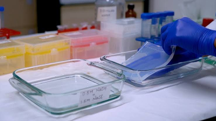
- After electrophoresis, carefully remove the gel and place it in a transfer buffer.
- Use capillary action or vacuum blotting techniques to transfer DNA from the gel onto a nylon or nitrocellulose membrane.
- Ensure that all fragments are transferred uniformly across the membrane surface.
- Crosslinking:
- Crosslink the transferred DNA to the membrane using UV light or heat treatment, which helps stabilize binding during subsequent steps.
Step 5: Prehybridization
- Blocking Non-Specific Binding Sites:
- Incubate the membrane in prehybridization buffer (containing blocking agents like salmon sperm DNA or denatured milk proteins) for several hours at an appropriate temperature (usually around 42°C).
Step 6: Probe Preparation
- Non-Radioactive Probe Labeling:
- Synthesize or obtain a probe that is complementary to your target sequence within B. subtilis. This probe can be labeled using biotin or digoxigenin, which allows for non-radioactive detection.
- Hybridization:
- Add labeled probe to hybridization buffer and incubate with the membrane overnight at an optimal temperature (typically around 42°C).
Step 7: Washing Steps
- Post-Hybridization Washes:
- Wash membranes multiple times with washing buffer at increasing stringency levels (lower salt concentrations) to remove non-specifically bound probes while retaining specific hybrids.
Step 8: Detection
- Non-Radioactive Detection Methods:
- Use enzyme-linked secondary antibodies that bind specifically to biotin or digoxigenin-labeled probes.
- Apply substrates for colorimetric detection (e.g., alkaline phosphatase substrates), allowing visualization of bound probes as colored bands on the membrane.
- Visualization:
- Capture images of membranes using imaging systems designed for chemiluminescent or colorimetric detection, enabling analysis of hybridized fragments corresponding to your target sequence in Bacillus subtilis.
By following these detailed steps, researchers can successfully perform Southern hybridization on Bacillus subtilis genomes utilizing non-radioactive detection methods, ensuring specificity and sensitivity in detecting desired genetic sequences.


