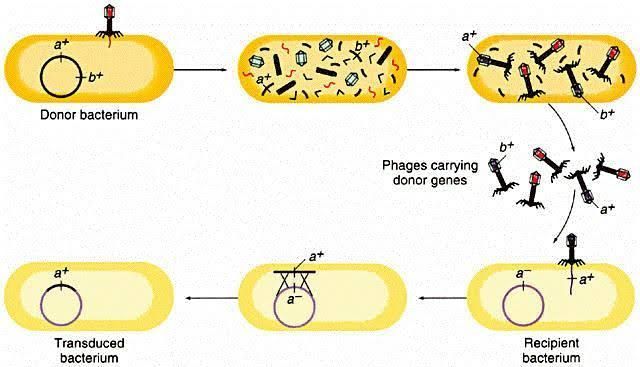
Genetic mapping using P1 transduction is a powerful method for transferring genetic material between bacterial strains, particularly in Escherichia coli (E. coli). This technique allows researchers to map the locations of genes on the bacterial chromosome by utilizing bacteriophage P1, which can carry segments of the host’s DNA during its lytic cycle. Below is a detailed step-by-step explanation of the procedures involved in genetic mapping via P1 transduction.
1. Preparation of Donor and Recipient Strains
The first step involves selecting appropriate donor and recipient strains of E. coli:
- Donor Strain: The donor strain should contain specific mutations or markers that are to be mapped. These mutations can be antibiotic resistance genes or other selectable traits.
- Recipient Strain: The recipient strain should ideally lack the mutations present in the donor strain and must be capable of homologous recombination, which is essential for integrating the transduced DNA into its genome.
2. Preparation of P1 Bacteriophage Lysate
To facilitate transduction, a lysate of bacteriophage P1 must be prepared:
- Grow the donor strain containing the desired mutation in a suitable medium until it reaches mid-log phase.
- Infect this culture with P1 bacteriophage at an appropriate multiplicity of infection (MOI), typically around 0.5 to ensure efficient infection without overwhelming the cells.
- Allow sufficient time for phage replication, usually several hours, until lysis occurs and phages are released into the medium.
- Collect and filter the lysate to remove bacterial debris, resulting in a concentrated solution of P1 bacteriophages.
3. Transduction Process
Once you have prepared your phage lysate, you can proceed with transduction:
- Inoculate the recipient strain with a small volume of the filtered phage lysate.
- Incubate this mixture under conditions that promote adsorption (e.g., at 37°C) for about 15–30 minutes.
- After incubation, add soft agar to allow plaque formation and further incubate to enable phage-mediated transfer of genetic material from donor to recipient.
4. Selection of Transductants
After allowing time for transduction:
- Plate the infected recipient cells on selective media that contains antibiotics corresponding to the resistance markers carried by the donor strain.
- Only those cells that successfully incorporated the antibiotic resistance gene through homologous recombination will survive on this selective medium.
5. Verification of Gene Transfer
To confirm successful transduction:
- Isolate colonies from selective plates and perform PCR or other molecular techniques to verify that they carry the desired mutation or marker from the donor strain.
- This verification often includes checking for antibiotic sensitivity/resistance patterns consistent with those expected from successful gene transfer.
6. Mapping Genes Using Cotransduction Frequencies
Once you have confirmed successful transductants:
- Perform cotransduction experiments where multiple markers are tested simultaneously to determine their relative positions on the chromosome.
- Calculate cotransduction frequencies; if two genes are frequently cotransduced together, they are likely located close to each other on the chromosome.
7. Data Analysis and Interpretation
Finally, analyze your data:
- Use statistical methods to interpret cotransduction frequencies and construct genetic maps based on these relationships.
- The closer two genes are on a chromosome, the higher their cotransduction frequency will be; this information can help refine gene order and distances between them.
Conclusion
P1 transduction is an effective method for genetic mapping in E. coli, allowing researchers to transfer specific genetic traits between strains efficiently while providing insights into gene location and linkage relationships within bacterial genomes. By following these steps—preparing strains, generating phage lysates, conducting transductions, selecting transductants, verifying gene transfers, performing cotransduction analysis, and interpreting results—scientists can create detailed genetic maps that enhance our understanding of microbial genetics.


