Step 1: Sample Preparation
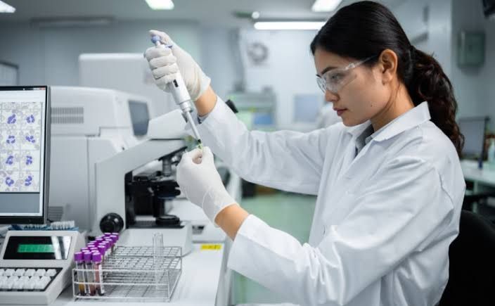
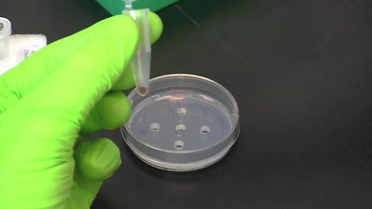
The first step in isolating DNA from bacteria involves preparing the bacterial sample. This can be done by culturing the bacteria in a suitable growth medium, such as LB broth, until they reach the logarithmic phase of growth. Once the culture is ready, it is harvested by centrifugation to collect the bacterial cells.
Step 2: Cell Lysis
To extract DNA, the bacterial cells must be lysed to release their contents. This can be achieved using various methods:
- Chemical Lysis: Detergents like sodium dodecyl sulfate (SDS) are commonly used to disrupt cell membranes. The detergent solubilizes lipids and proteins, facilitating the release of DNA into solution.
- Enzymatic Lysis: Enzymes such as lysozyme can be employed to break down the peptidoglycan layer of Gram-positive bacteria. This method is particularly effective for tougher cell walls.
- Physical Methods: Mechanical disruption techniques like bead beating or sonication can also be used to physically break open bacterial cells.
After lysis, the mixture contains cellular debris along with released DNA.
Step 3: Removal of Cellular Debris
Following lysis, it is crucial to remove cellular debris and contaminants that could interfere with downstream applications. This can be accomplished through:
- Centrifugation: The lysate is centrifuged at high speed to pellet insoluble materials. The supernatant containing the DNA is carefully collected.
- Filtration: Alternatively, filtration methods can be used to separate debris from the lysate.
Step 4: DNA Purification
Once the cellular debris has been removed, the next step is purifying the DNA. Common methods include:
- Silica-Based Extraction: In this method, chaotropic salts (like guanidine hydrochloride) are added to promote binding of DNA to silica membranes or beads under high-salt conditions. After binding, impurities are washed away with ethanol-based solutions before eluting pure DNA in a low-salt buffer or nuclease-free water.
- Phenol-Chloroform Extraction: This traditional method involves adding phenol-chloroform to separate nucleic acids from proteins and lipids based on solubility differences. After mixing and centrifuging, DNA partitions into the aqueous phase while proteins remain in the organic phase.
Step 5: Quantification and Quality Assessment
After purification, it’s essential to quantify and assess the quality of isolated DNA. Techniques include:
- Spectrophotometry: Measuring absorbance at 260 nm provides an estimate of DNA concentration; an A260/A280 ratio around 1.8 indicates good purity.
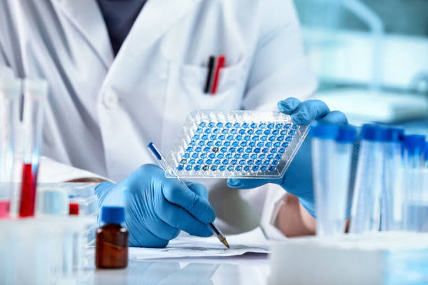
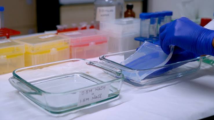
- Gel Electrophoresis: Running a sample on an agarose gel allows visualization of DNA integrity; intact bands indicate high-quality genomic DNA.
Step 6: Amplification of Specific Gene Sequence
With purified bacterial DNA in hand, specific gene sequences can now be amplified using Polymerase Chain Reaction (PCR). The steps involved are:
- Design Primers: Primers specific to the target gene sequence must be designed. These short sequences flank the region of interest and are critical for successful amplification.
- Prepare PCR Reaction Mix:
- Include template DNA (the purified bacterial genomic DNA).
- Add primers (forward and reverse).
- Include deoxynucleoside triphosphates (dNTPs), which serve as building blocks for new DNA strands.
- Use a thermostable polymerase enzyme (like Taq polymerase) that withstands high temperatures during PCR cycles.
- Add a buffer solution that maintains optimal pH and ionic strength for enzyme activity.
- Thermal Cycling:
- Denaturation (92–95°C): Heat denatures double-stranded DNA into single strands.
- Annealing (50–70°C): Lower temperature allows primers to bind specifically to their complementary sequences on single-stranded templates.
- Extension (72°C): The polymerase synthesizes new strands by adding dNTPs complementary to each template strand.
These steps are repeated for 25–40 cycles depending on desired yield.
- Post-PCR Analysis: After amplification, products can be analyzed via gel electrophoresis to confirm successful amplification of the target gene sequence by comparing band sizes against a molecular weight marker.
By following these detailed steps for isolation and amplification, researchers can effectively obtain high-quality bacterial genomic DNA and amplify specific gene sequences for further analysis or applications in molecular biology research.


