Protein Motif and Domain Architecture
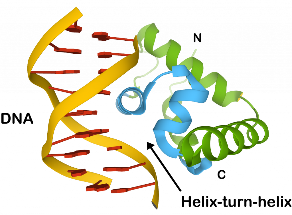
Protein motifs are short, conserved sequences of amino acids that are associated with specific functions or structural features in proteins. These motifs can be part of larger protein domains, which are distinct functional and structural units within a protein. The architecture of a protein is determined by the arrangement and interaction of these motifs and domains, which ultimately influence the protein’s function.
Sequence-Structure Mapping and Protein Folding
The relationship between a protein’s sequence (the order of amino acids) and its three-dimensional structure is crucial for understanding how proteins fold. Protein folding is driven by various forces and interactions, including:
- Hydrophobic interactions: Non-polar side chains tend to avoid water, driving them to the interior of the protein.
- Hydrogen bonds: These occur between polar side chains or backbone atoms, stabilizing secondary structures like alpha helices and beta sheets.
- Ionic interactions: Charged side chains can form salt bridges that stabilize the folded structure.
- Van der Waals forces: Weak attractions between all atoms contribute to overall stability.
Understanding this mapping helps in predicting how changes in sequence can affect structure and function.
Protein Sequence Predictions
Protein sequence predictions can be categorized into three main approaches:
- Ab initio methods: These rely on physical principles and statistical models to predict protein structure from scratch without using known structures as templates.
- Homology-based methods: These use known structures of homologous proteins (proteins with similar sequences) as templates to predict the structure of an unknown protein based on sequence similarity.
- Threading methods: These involve aligning a target sequence onto a library of known structures to find the best fit, even if there is low sequence similarity.
Each method has its strengths and weaknesses depending on the availability of data and computational resources.
Protein Identification and Quantification
Several techniques are used for identifying and quantifying proteins:
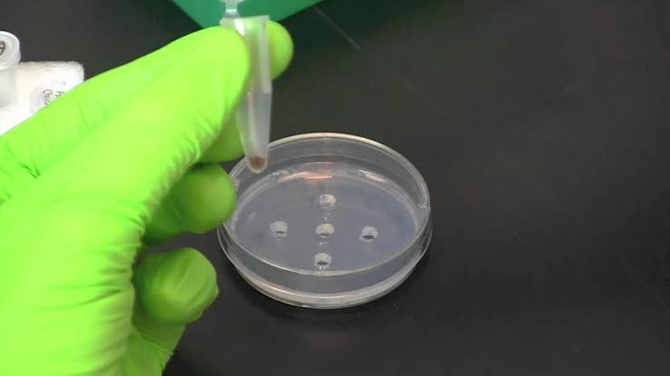
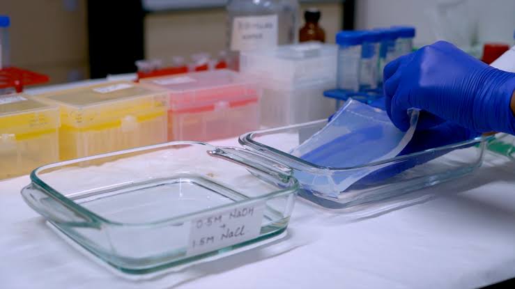
- 2D Gel Electrophoresis: This technique separates proteins based on their isoelectric point (pI) and molecular weight, allowing for visualization of complex mixtures.
- Mass Spectrometry (MALDI-TOF): This method measures the mass-to-charge ratio of ionized particles, providing precise identification of proteins based on their mass.
- Other Arrays: Techniques such as protein microarrays allow for simultaneous analysis of multiple proteins through binding assays.
- Yeast Two-Hybrid System: This genetic method detects interactions between two proteins by reconstituting a functional transcription factor in yeast cells.
- ICAT (Isotope-Coded Affinity Tagging): This technique allows for quantitative comparison of protein expression levels across different samples by tagging proteins with isotopes.
Protein-DNA Recognition
The recognition between proteins and DNA involves specific models and algorithms that help predict how proteins bind to DNA sequences:
- Structural models: These provide insights into how specific amino acid residues interact with DNA bases through hydrogen bonding, hydrophobic interactions, or electrostatic forces.
- Algorithms: Computational tools analyze DNA sequences to identify potential binding sites based on known binding patterns from characterized protein-DNA complexes.
These models are essential for understanding gene regulation mechanisms.
In summary, understanding protein motifs, folding mechanisms, prediction methods, identification techniques, and recognition processes provides a comprehensive view of proteomics as it relates to biological functions.


