Necessary Precautions While Working with RNA
When working with RNA, it is crucial to take specific precautions to prevent degradation and contamination. Here are the key steps:
- Use RNase-Free Equipment: All equipment, including pipettes, tubes, and tips, should be certified RNase-free. This helps prevent contamination from ribonucleases that can degrade RNA.
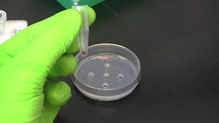
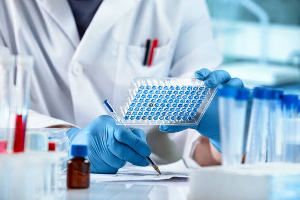
- Wear Gloves: Always wear gloves when handling RNA or any materials that will come into contact with RNA. This minimizes the risk of introducing RNases from skin or other surfaces.
- Work in a Clean Environment: Use a clean bench or laminar flow hood to minimize exposure to airborne contaminants. Regularly clean surfaces with RNase decontamination solutions.
- Keep Samples on Ice: During isolation and manipulation of RNA, keep samples on ice to slow down enzymatic activity that could lead to degradation.
- Use Appropriate Reagents: Employ reagents specifically designed for RNA work, such as those containing stabilizers that protect RNA from degradation.
Isolation of RNA from Clinical Samples
The isolation of RNA from clinical samples involves several critical steps:
- Sample Collection: Collect samples in RNase-free tubes and immediately process them or store them at -80°C if not processed right away.
- Cell Lysis: Use a lysis buffer containing chaotropic agents (like guanidine thiocyanate) to disrupt cells and release RNA into solution.
- Phase Separation: Add phenol-chloroform to separate the aqueous phase (which contains RNA) from the organic phase (which contains proteins and DNA).
- Precipitation: Precipitate the RNA by adding isopropanol or ethanol, followed by centrifugation to collect the RNA pellet.
- Washing and Resuspension: Wash the pellet with 70% ethanol to remove impurities and then resuspend it in RNase-free water or buffer.
Preparation of cDNA
Complementary DNA (cDNA) synthesis involves reverse transcription of mRNA:
- Template Preparation: Start with purified mRNA isolated from your sample.
- Reverse Transcription Reaction:
- Mix mRNA with reverse transcriptase enzyme, primers (oligo(dT) or random hexamers), dNTPs, and buffer.
- Incubate under optimal conditions for reverse transcriptase activity (usually around 42°C).
- Termination of Reaction: Heat inactivate the reverse transcriptase by incubating at 70°C for a few minutes.
- Storage: Store cDNA at -20°C for future use in PCR or other applications.
Northern Blotting and Reverse-Transcription PCR
(a) Northern Blotting
Northern blotting is used for detecting specific RNA sequences:
- Gel Electrophoresis: Separate RNA samples by size using agarose gel electrophoresis.
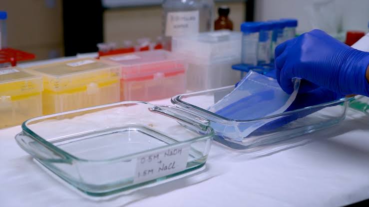
- Transfer to Membrane: Transfer the separated RNA onto a nylon or nitrocellulose membrane using capillary action or electroblotting.
- Hybridization:
- Incubate the membrane with labeled probes complementary to target sequences.
- Wash off unbound probes under stringent conditions.
- Detection: Visualize bound probes using autoradiography or chemiluminescence methods.
(b) Reverse-Transcription PCR (RT-PCR)
RT-PCR combines reverse transcription and PCR amplification:
- Reverse Transcription Step: Convert mRNA into cDNA as previously described.
- PCR Amplification Step:
- Use specific primers for target genes.
- Perform thermal cycling (denaturation, annealing, extension) to amplify cDNA into detectable products.
- Analysis of Products: Analyze PCR products via gel electrophoresis or quantitative methods like qPCR for expression levels.
Detection of Viral Pathogens
HCV, HIV, Influenza Virus Detection
Detection methods include:
- Molecular Techniques (PCR):
- Utilize specific primers targeting viral genomes for amplification.
- Real-time PCR can quantify viral load effectively.
- Serological Tests:
- Rapid Antigen Tests:
- These tests provide quick results but may have lower sensitivity compared to molecular methods.
Genotyping of Viral Strains (HCV)
Genotyping involves determining the strain of HCV present in a sample:
- Sequencing Methods:
- Amplify regions of interest through PCR followed by Sanger sequencing or next-generation sequencing techniques.
- Phylogenetic Analysis:
- Compare sequences against known databases to classify strains based on genetic similarities and differences.
Detection of Oncogene Expression
Her2/neu, BRCA1/BRCA2, N-myc Expression Detection
Methods include:
- Quantitative RT-PCR (qRT-PCR):
- Measure expression levels using specific primers for each oncogene.
- In Situ Hybridization (ISH):
- Localize gene expression within tissue sections using labeled probes that bind specifically to target mRNAs.
Quantitative Expression of Fusion Transcripts
BCR-ABL, PML-RARA, TEL-AML Detection
Quantitative detection involves:
- Specific Primers Design:
- Design primers that flank fusion junctions unique to each transcript.
- Real-Time PCR Analysis:
- Perform qPCR using these primers alongside reference genes for normalization purposes.
- Data Interpretation:
- Analyze Ct values relative to controls to determine expression levels quantitatively across different samples.
In summary, working with RNA requires meticulous attention to detail regarding contamination prevention during isolation processes while employing various techniques such as Northern blotting and RT-PCR for downstream applications like pathogen detection and oncogene expression analysis.


