Formation and Folding of the Heart Tube
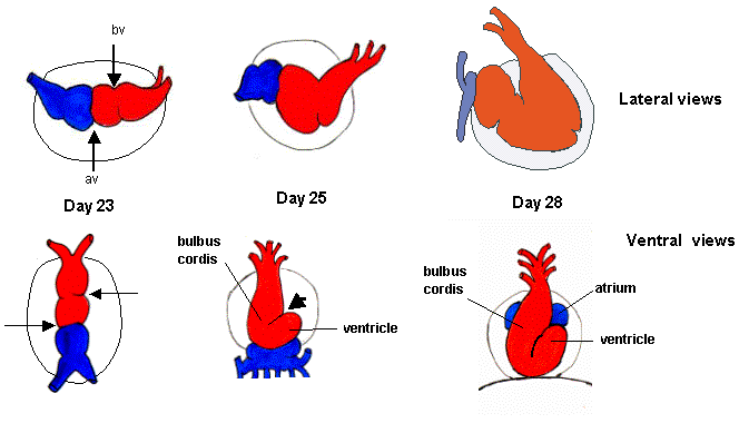
The heart tube is the precursor to the mature heart and forms during early embryonic development. The process begins around the third week of gestation, primarily through a series of complex interactions involving mesodermal cells.
Primary Formation of the Heart Tube
- Mesodermal Development: The heart originates from mesodermal progenitor cells located in the lateral plate mesoderm. These cells migrate to form two distinct regions known as the cardiogenic area.
- Formation of Cardiac Primordium: As development progresses, these progenitor cells differentiate into cardiac myocytes and endothelial cells, forming a structure called the cardiac primordium.
- Heart Tube Formation: By day 21 post-fertilization, these structures fuse to form a linear heart tube. This tube consists of three layers:
- Endocardium: The inner layer that lines the heart chambers.
- Myocardium: The middle muscular layer responsible for contraction.
- Epicardium: The outer layer that provides protection and contains coronary vessels.
- Folding of the Heart Tube: Following its formation, the heart tube undergoes a process called looping or folding, which occurs around day 28. This involves:
- The bending of the tube at specific points to create distinct regions that will develop into various chambers.
- The atrial region moves cranially while the ventricle region shifts caudally, resulting in a more complex structure.
Formation of Different Chambers of the Heart
The looping process leads to further differentiation into specific chambers:
- Atria and Ventricles Development:
- Initially, there are no distinct atria or ventricles; however, as looping continues, bulges form that will become future chambers.
- The primitive ventricle becomes more prominent while an outflow tract develops into what will be the future aorta and pulmonary artery.
- Septation Process:
- Septation refers to the division of single chambers into left and right sides through septa formation.
- Atrial septation occurs via two key structures:
- The septum primum grows downward from the roof of the atrium toward the endocardial cushions.
- Subsequently, septum secundum forms alongside it but does not completely close off; this creates openings (foramen ovale) allowing blood flow between atria during fetal life.
- Ventricular septation involves growth from both sides towards each other until they fuse in the midline, creating left and right ventricles.
- Valvular Development:
- As chambers form, valves develop from endocardial cushions that grow into these spaces to ensure unidirectional blood flow once circulation begins.
Establishment of Fetal Circulation
Fetal circulation is characterized by unique adaptations that facilitate oxygen delivery from maternal blood:
- Key Structures in Fetal Circulation:
- Placenta: Acts as an organ for gas exchange; oxygenated blood returns to fetus via umbilical vein.
- Ductus Venosus: Bypasses liver circulation directly into inferior vena cava.
- Foramen Ovale: Allows blood flow between right and left atria.
- Ductus Arteriosus: Connects pulmonary artery to descending aorta, bypassing non-functioning lungs.
- Hemodynamics During Fetal Life:
- Blood flows preferentially through these shunts due to pressure differences; right atrial pressure is higher than left atrial pressure facilitating foramen ovale function.
- Systemic circulation is established with lower resistance compared to pulmonary circulation due to non-ventilated lungs.
Cardiovascular Changes After Birth
Upon birth, significant cardiovascular changes occur:
- Closure of Shunts:
- Increased oxygen levels lead to vasodilation and closure of ductus arteriosus within hours after birth.
- Foramen ovale closes due to increased left atrial pressure as lungs expand with air intake.
- Transitioning Circulation Pathways:
- Blood flow patterns shift dramatically; systemic resistance increases while pulmonary resistance decreases leading to normal adult circulation patterns.
- Adaptations in Cardiac Functionality:
- The heart must adapt quickly to new hemodynamic conditions; myocardial remodeling occurs over time as it adjusts from fetal patterns to adult functionality.
Causes of Major Congenital Malformations
Congenital malformations can arise during any stage of cardiac development:
- Genetic Factors:
- Chromosomal abnormalities such as Down syndrome (Trisomy 21) are associated with congenital heart defects like atrioventricular septal defects (AVSD).
- Environmental Influences:
- Teratogens such as alcohol (fetal alcohol syndrome) or certain medications can disrupt normal cardiac development leading to structural anomalies like ventricular septal defects (VSD).
- Clinical Implications:
- Congenital malformations can lead to significant morbidity and mortality if not diagnosed early; management may require surgical intervention or lifelong monitoring depending on severity.
In summary, understanding these developmental processes is crucial for recognizing potential congenital issues early on and providing appropriate care strategies for affected individuals.


