Metabolism of Blood Cells
1. Reticulocytes
Reticulocytes are immature red blood cells (RBCs) that are produced in the bone marrow and released into the bloodstream. Their metabolism is primarily focused on hemoglobin synthesis and energy production. They contain ribosomal RNA, which gives them a reticular appearance when stained. The metabolic processes include:
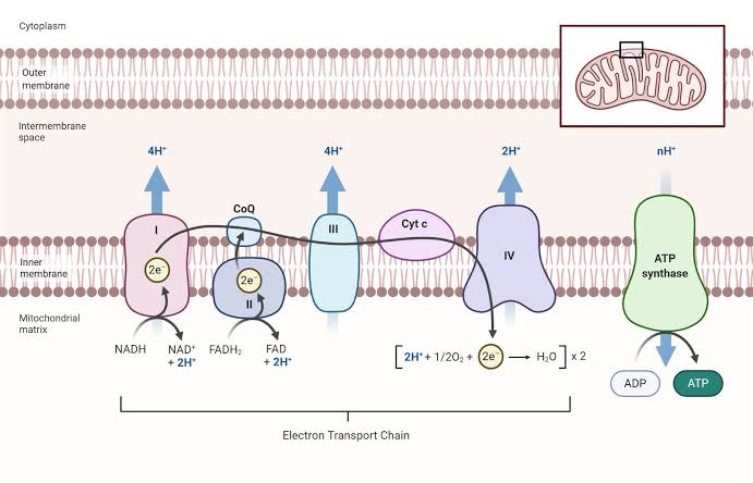
- Glycolysis: Reticulocytes rely heavily on anaerobic glycolysis for ATP production, as they lack mitochondria.
- Hemoglobin Synthesis: They synthesize hemoglobin until they mature into erythrocytes, utilizing iron and porphyrin for heme production.
- RNA Degradation: As they mature, reticulocytes gradually degrade their RNA content.
2. Erythrocytes
Mature erythrocytes have a lifespan of about 120 days and primarily function to transport oxygen and carbon dioxide. Their metabolism includes:
- Anaerobic Glycolysis: Erythrocytes depend on glycolysis for ATP generation since they lack mitochondria.
- Pentose Phosphate Pathway (PPP): This pathway is crucial for generating NADPH, which protects against oxidative damage by maintaining glutathione levels.
- Hemoglobin Function: Erythrocytes utilize hemoglobin to bind oxygen in the lungs and release it in tissues.
3. Leucocytes
Leucocytes (white blood cells) are involved in immune responses and have diverse metabolic pathways depending on their type (e.g., neutrophils, lymphocytes). Key metabolic features include:
- Aerobic Respiration: Some leucocytes can perform aerobic respiration to generate ATP.
- Reactive Oxygen Species (ROS) Production: Neutrophils produce ROS during phagocytosis to kill pathogens.
- Cytokine Production: Lymphocytes metabolize nutrients to produce cytokines that regulate immune responses.
4. Platelets
Platelets are cell fragments involved in hemostasis. Their metabolism includes:
- Glycolysis: Like other blood cells, platelets rely on glycolysis for energy.
- Activation Pathways: Upon activation, platelets undergo shape change and granule release, which requires ATP.
- Arachidonic Acid Metabolism: Platelets convert arachidonic acid into thromboxane A2, promoting vasoconstriction and platelet aggregation.
Blood Coagulation Factors
Blood coagulation involves a series of proteins known as coagulation factors. These factors are typically designated by Roman numerals I through XIII (excluding factor VI). Here’s a list of key factors along with their properties:
- Factor I (Fibrinogen): Soluble plasma protein converted to fibrin during clot formation.
- Factor II (Prothrombin): Precursor to thrombin; requires vitamin K for synthesis.
- Factor III (Tissue Factor): Initiates extrinsic pathway; released from damaged tissues.
- Factor IV (Calcium ions): Essential cofactor in multiple steps of coagulation cascade.
- Factor V (Proaccelerin): Acts as a cofactor for prothrombinase complex formation.
- Factor VII (Proconvertin): Activates factor X in the presence of tissue factor.
- Factor VIII (Antihemophilic Factor A): Cofactor for factor IX; deficiency leads to hemophilia A.
- Factor IX (Christmas Factor): Activates factor X; deficiency leads to hemophilia B.
- Factor X (Stuart-Prower Factor): Converts prothrombin to thrombin; central role in coagulation cascade.
- Factor XI (Plasma Thromboplastin Antecedent): Activates factor IX; involved in intrinsic pathway activation.
- Factor XII (Hageman Factor): Initiates intrinsic pathway upon contact with negatively charged surfaces.
- Factor XIII (Fibrin-stabilizing Factor): Cross-links fibrin strands to stabilize the clot.
Extrinsic and Intrinsic Pathways of Coagulation
Coagulation occurs via two main pathways that converge at factor X activation:
Extrinsic Pathway
The extrinsic pathway is initiated by tissue injury leading to exposure of tissue factor:
- Tissue factor binds with circulating factor VIIa in the presence of calcium ions.
- This complex activates factor X, leading to thrombin generation.
This pathway is rapid and is primarily responsible for immediate clot formation following vascular injury.
Intrinsic Pathway
The intrinsic pathway is activated by damage to the vessel wall or exposure of collagen:
- Factor XII comes into contact with negatively charged surfaces, activating it to XIIa.
- Activated XIIa then activates XIa, which subsequently activates IXa in conjunction with its cofactor VIIIa.
- Finally, this complex activates factor X.
The intrinsic pathway is slower but amplifies the coagulation response initiated by the extrinsic pathway.
Inhibition of Clotting by Oxalate, Fluoride, EDTA
These anticoagulants inhibit clotting through different mechanisms:
- Oxalate:
- Oxalate chelates calcium ions necessary for various steps in the coagulation cascade, particularly affecting factors II, VII, IX, and X.
- Fluoride:
- Fluoride also chelates calcium ions but additionally inhibits enzymes involved in glycolysis within platelets, impairing their function.
- EDTA (Ethylenediaminetetraacetic Acid):
- EDTA binds calcium ions effectively preventing their availability for coagulation reactions involving several factors including prothrombin activation.
Function of Plasminogen and Clot Lysis
Plasminogen is an inactive precursor that plays a crucial role in fibrinolysis—the process that dissolves clots:
- Plasminogen is incorporated into forming clots where it remains inactive until activated by tissue plasminogen activator (tPA) or urokinase-type plasminogen activator (uPA).
- Once activated into plasmin, it digests fibrin strands within the clot leading to its dissolution—a critical step ensuring normal blood flow resumes after healing.
Defects Related to Hemostasis
Defects can occur at various levels affecting hemostasis:
1. Platelet Defects
- Thrombocytopenia or dysfunctional platelets can lead to bleeding disorders such as Glanzmann’s thrombasthenia or Bernard-Soulier syndrome.
2. Plasma Defects
- Deficiencies or dysfunctions of coagulation factors can result from genetic conditions like hemophilia or von Willebrand disease affecting clotting ability.
3. Vessel Wall Defects
- Conditions such as Ehlers-Danlos syndrome affect collagen integrity within vessel walls leading to increased fragility and bleeding tendencies.
Biochemical Explanation of Hemorrhagic Disease
Hemorrhagic diseases arise from deficiencies or dysfunctions within any component involved in hemostasis—platelets, plasma proteins or vessel walls—leading to excessive bleeding tendencies due to inadequate clot formation or stability:
- Genetic disorders like hemophilia result from specific factor deficiencies disrupting normal coagulation cascades leading directly to bleeding episodes.
- Acquired conditions such as liver disease impair synthesis of multiple clotting factors due to reduced hepatic function resulting in similar bleeding risks.
In summary, understanding these components provides insight into both normal hemostatic processes and pathological states associated with bleeding disorders.


