Common Causes of Lower Gastro-Intestinal Bleeding
Lower gastrointestinal bleeding (LGIB) can arise from various conditions affecting the colon, rectum, and anus. The common causes include:
- Diverticular Disease: Diverticulosis can lead to diverticulitis or diverticular bleeding.
- Hemorrhoids: Swollen veins in the lower rectum or anus can cause bright red blood during bowel movements.
- Colorectal Polyps: Benign growths that may bleed, especially if they are large or dysplastic.
- Colorectal Cancer: Malignant tumors can cause bleeding, often associated with changes in bowel habits.
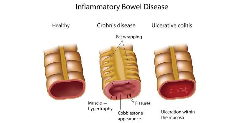
- Inflammatory Bowel Disease (IBD): Includes ulcerative colitis and Crohn’s disease, both of which can cause significant bleeding.
- Infectious Colitis: Infections from bacteria, viruses, or parasites can lead to inflammation and bleeding.
- Angiodysplasia: Abnormal blood vessels in the gastrointestinal tract that can rupture and bleed.
- Ischemic Colitis: Reduced blood flow to the colon leading to tissue damage and potential bleeding.
Analyzing Patient History for LGIB
When assessing a patient with suspected LGIB, it is crucial to gather a comprehensive history:
- Symptoms:
- Onset of bleeding (acute vs chronic)
- Nature of the blood (bright red vs dark/maroon)
- Associated symptoms (abdominal pain, diarrhea, weight loss)
- Medical History:
- Previous episodes of GI bleeding
- History of IBD, colorectal cancer, or polyps
- Use of medications (NSAIDs, anticoagulants)
- Family History:
- Family history of colorectal cancer or polyps
- Social History:
- Dietary habits
- Alcohol use
- Smoking status
By interpreting these findings, one may identify specific causes such as hemorrhoids for bright red blood on toilet paper or diverticular disease for maroon-colored stools.
Management Strategy for LGIB
The management strategy for a patient suffering from lower gastrointestinal bleeding includes:
- Initial Assessment:
- Stabilization of vital signs
- Establishing IV access for fluid resuscitation if necessary
- Diagnostic Evaluation:
- Perform a digital rectal examination to assess for hemorrhoids or masses.
- Order laboratory tests including CBC to evaluate hemoglobin levels.
- Endoscopy:
- Depending on the severity and source suspected, perform a colonoscopy for direct visualization and possible intervention.
- Surgical Intervention:
- If endoscopic measures fail and there is ongoing significant bleeding, surgical intervention may be required.
- Follow-Up Care:
- Monitor hemoglobin levels post-management and provide appropriate follow-up care based on findings.
Diagnosing Inflammatory Bowel Disease
Diagnosis of inflammatory bowel disease (IBD), including ulcerative colitis and Crohn’s disease involves:
- History Taking:
- Symptoms such as diarrhea (often bloody), abdominal pain, urgency, weight loss.
- Physical Examination:
- Abdominal tenderness; check for perianal disease in Crohn’s patients.
- Differentiation Between Types:
- Ulcerative colitis typically presents with continuous lesions starting from the rectum.
- Crohn’s disease may present with skip lesions affecting any part of the GI tract.
Ordering Investigations for IBD
Appropriate investigations include:
- Laboratory Tests:
- Complete blood count (CBC) to check for anemia.
- C-reactive protein (CRP) and erythrocyte sedimentation rate (ESR) to assess inflammation.
- Imaging Studies:
- Colonoscopy with biopsy is essential for diagnosis; it allows direct visualization and histological assessment.
- Radiological Imaging:
- MRI enterography or CT enterography may be used particularly in suspected Crohn’s disease to assess small bowel involvement.
Management Plan for IBD
A comprehensive management plan includes:
- Medications:
- Aminosalicylates (e.g., mesalamine) for mild cases.
- Corticosteroids for moderate-severe flares.
- Immunomodulators (e.g., azathioprine) or biologics (e.g., infliximab) for maintenance therapy.
- Nutritional Support:
- Dietary modifications tailored to individual tolerance; consider nutritional supplements if malnutrition is present.
- Surgery Consideration:
- Surgical options may be necessary in cases of complications like strictures or abscesses in Crohn’s disease; colectomy may be indicated in severe ulcerative colitis cases unresponsive to medical therapy.
- Regular Monitoring & Follow-Up Care – Regular follow-ups with gastroenterology are essential to monitor disease activity and adjust treatment as needed.
Stoma Formation Principles
When considering stoma formation following surgery:
- Site Selection – The stoma should ideally be placed on the right side of the abdomen where there is adequate skin surface area away from bony prominences.
- Type of Stoma – An ileostomy is typically formed when the entire colon is removed; a colostomy may be performed when part of the colon remains intact depending on pathology involved.
Complications of Stoma
Complications associated with stomas include:
- Skin irritation around the stoma site due to leakage.
- Stenosis at the stoma site leading to obstruction.
- Prolapse where the stoma protrudes excessively outside the abdominal wall.
- Parastomal hernia where abdominal contents protrude through weakened abdominal muscles near the stoma site.
Counseling Regarding Stoma Formation
Counseling should encompass:
- Explanation about what a stoma is and why it is necessary post-surgery due to carcinoma rectum resection.
- Discussion about lifestyle changes including dietary adjustments post-stoma formation.
- Instruction on proper stoma care techniques including cleaning and changing bags effectively while addressing concerns regarding body image issues related to having a stoma.
Types and Classification of Intestinal Fistulas
Intestinal fistulas can be classified into several types based on their origin:
- Enteric Fistulas: Between two segments of intestine.
- Entero-cutaneous Fistulas: Between intestine and skin surface.
- Entero-vaginal Fistulas: Between intestine and vagina.
- Entero-urinary Fistulas: Between intestine and urinary tract structures.
Management Options
Management strategies depend on fistula type but generally include:
- Conservative Management: Nutritional support via TPN if needed; maintaining skin integrity around fistula sites; monitoring output closely until spontaneous closure occurs if possible.
- Surgical Intervention: May be required if conservative measures fail after an appropriate duration or if complications arise such as infection or obstruction related directly due to fistula presence.


