Normal Histological Presentation of Skeletal Muscle
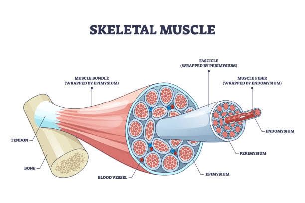
Skeletal muscle is characterized by its striated appearance under the microscope, which is due to the organized arrangement of myofibrils. The histological presentation includes:
- Muscle Fibers: Skeletal muscle fibers are long, cylindrical cells that can vary in length and diameter. They are multinucleated, with nuclei located at the periphery of the cell.
- Striations: The striations are a result of the repeating units called sarcomeres, which contain thick (myosin) and thin (actin) filaments. This organization gives skeletal muscle its characteristic banding pattern.
- Connective Tissue: Surrounding each muscle fiber is a layer of connective tissue called endomysium. Groups of muscle fibers are bundled together into fascicles, which are surrounded by perimysium. The entire muscle is encased in epimysium.
- Blood Supply and Nerves: Skeletal muscles have a rich blood supply and innervation, with capillaries running between the fibers and motor neurons connecting to each fiber at neuromuscular junctions.
- Satellite Cells: These are stem cells located between the basal lamina and the sarcolemma of muscle fibers, playing a crucial role in muscle repair and regeneration.
Classification of Diseases Related to Skeletal Muscle
Diseases affecting skeletal muscle can be classified into several categories:
- Inherited Myopathies:
- Duchenne Muscular Dystrophy (DMD): A genetic disorder caused by mutations in the dystrophin gene.
- Becker Muscular Dystrophy: A milder form of muscular dystrophy also related to dystrophin mutations.
- Myotonic Dystrophy: Characterized by prolonged muscle contractions and weakness.
- Acquired Myopathies:
- Inflammatory Myopathies: Such as polymyositis and dermatomyositis, where the immune system attacks muscle tissue.
- Infectious Myopathies: Caused by infections like viral myositis or parasitic infections such as trichinosis.
- Endocrine Myopathies: Resulting from hormonal imbalances, such as hypothyroidism or Cushing’s syndrome.
- Metabolic Myopathies:
- Disorders affecting energy metabolism within muscles, such as glycogen storage diseases or mitochondrial myopathies.
- Toxic Myopathies:
- Caused by exposure to toxins or drugs, including alcohol-related myopathy or statin-induced myopathy.
- Neuromuscular Junction Disorders:
- Conditions like myasthenia gravis that affect communication between nerves and muscles.
Pathophysiological Patterns for Skeletal Muscle Pathologies
The pathophysiology of skeletal muscle diseases often involves:
- Degeneration and Regeneration: In many myopathies, there is an initial phase of degeneration where muscle fibers undergo necrosis followed by attempts at regeneration mediated by satellite cells.
- Inflammation: Inflammatory myopathies show infiltration of immune cells leading to further damage to muscle fibers.
- Fibrosis: Chronic injury often leads to fibrosis where normal muscle tissue is replaced with scar tissue, impairing function.
- Metabolic Dysfunction: In metabolic disorders, there may be an inability to utilize energy substrates effectively leading to exercise intolerance or weakness during exertion.
- Neuromuscular Transmission Failure: In conditions affecting neuromuscular junctions, there is impaired transmission leading to weakness that typically worsens with activity (fatigable weakness).
Management Plan for Various Muscle Pathologies
Management strategies depend on the specific pathology but generally include:
- Physical Therapy: Essential for maintaining strength and function in various myopathies; tailored exercise programs help prevent contractures and maintain mobility.
- Medications:
- Corticosteroids: Often used in inflammatory myopathies to reduce inflammation.
- Immunosuppressants: Such as azathioprine or methotrexate for autoimmune conditions.
- Enzyme Replacement Therapy: For certain metabolic disorders like Pompe disease.
- Nutritional Support: Important for metabolic myopathies; dietary adjustments may be necessary based on specific enzyme deficiencies (e.g., high-protein diets for certain glycogen storage diseases).
- Surgical Interventions: May be indicated for severe contractures or deformities resulting from chronic weakness or injury.
- Genetic Counseling and Supportive Care: For inherited conditions like muscular dystrophies; providing resources for families affected by genetic disorders is crucial.
- Monitoring for Complications: Regular follow-ups are necessary to manage potential complications such as respiratory failure in severe cases or cardiac involvement in certain muscular dystrophies.
In conclusion, understanding skeletal muscle histology aids in recognizing various pathologies associated with it while guiding appropriate management strategies tailored to individual needs based on specific diagnoses.


