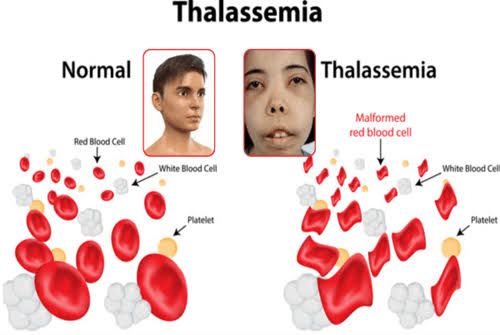
Thalassemia syndromes are a group of inherited blood disorders characterized by reduced or absent synthesis of one or more globin chains that make up hemoglobin. They can lead to anemia and various complications depending on the severity and type of thalassemia.
Types of Thalassemia
Thalassemias are primarily classified into two major types based on which globin chain is affected:
- Alpha Thalassemia: This type results from mutations in the alpha-globin genes located on chromosome 16. There are four alpha-globin genes, and the severity of alpha thalassemia depends on how many of these genes are mutated:
- Silent Carrier: One gene is mutated; usually asymptomatic.
- Alpha Thalassemia Trait (Minor): Two genes are mutated; mild anemia may occur.
- Hemoglobin H Disease: Three genes are mutated; moderate to severe anemia occurs.
- Alpha Thalassemia Major (Hydrops Fetalis): All four genes are mutated; this condition is typically fatal before or shortly after birth.
- Beta Thalassemia: This type arises from mutations in the beta-globin gene located on chromosome 11. The severity also varies based on the number of affected alleles:
- Beta Thalassemia Minor (Trait): One gene is mutated; often asymptomatic with mild anemia.
- Beta Thalassemia Intermedia: Both genes have mutations, but some beta-globin production remains; moderate anemia occurs.
- Beta Thalassemia Major (Cooley’s Anemia): Both genes are severely mutated, leading to little or no beta-globin production; severe anemia develops early in life.
Types of Mutations
Mutations causing thalassemias can be classified into several categories:
- Point Mutations: These involve a single nucleotide change in the DNA sequence, which can lead to an amino acid substitution or create a premature stop codon.
- Deletions: Portions of the gene may be deleted, leading to loss of function. This is particularly common in alpha thalassemias where entire alpha-globin genes may be deleted.
- Insertions: Additional nucleotides may be inserted into the gene, potentially disrupting normal protein synthesis.
- Splice Site Mutations: Changes at the boundaries between exons and introns can affect mRNA splicing, resulting in abnormal protein products.
Pathogenesis of Thalassemia Syndromes
The pathogenesis of thalassemic syndromes involves several interconnected processes:
- Imbalanced Globin Chain Production: In thalassemias, there is an imbalance between alpha and beta globin chain production. For instance, in beta thalassemia major, there is insufficient beta globin produced relative to alpha globin, leading to excess free alpha chains.
- Ineffective Erythropoiesis: The excess unpaired globin chains precipitate within erythroid precursors in the bone marrow, leading to apoptosis (programmed cell death) and ineffective erythropoiesis. This results in reduced red blood cell production.
- Hemolysis: The imbalance also causes increased destruction of mature red blood cells due to membrane instability caused by excess unpaired chains.
- Compensatory Mechanisms: The body attempts to compensate for anemia through increased erythropoietin production and extramedullary hematopoiesis (blood cell formation outside the bone marrow), often leading to splenomegaly and other complications.
- Iron Overload: Patients with chronic hemolytic anemia often require multiple blood transfusions for management, which can lead to secondary iron overload due to excessive iron absorption from the gut and accumulation in organs such as the liver and heart.
Morphological Features on Peripheral Blood Smear
When examining a peripheral blood smear from patients with thalassemia syndromes, several characteristic features can be observed:
- Microcytic Hypochromic Anemia: Red blood cells appear smaller than normal (microcytic) and have less color (hypochromic) due to decreased hemoglobin content.
- Target Cells: These cells have a bullseye appearance due to an abnormal distribution of hemoglobin within them.
- Basophilic Stippling: This refers to small blue granules seen within red blood cells due to residual RNA or ribosomal material.
- Anisocytosis and Poikilocytosis: There is variability in red blood cell size (anisocytosis) and shape (poikilocytosis).
- Increased Reticulocytes: A higher number of immature red blood cells may be present as a response to anemia.
- Nucleated Red Blood Cells (NRBCs): In severe cases like beta thalassemia major, nucleated red blood cells may appear in circulation due to extramedullary hematopoiesis.
In summary, understanding thalassemia syndromes involves recognizing their genetic basis through different types of mutations, comprehending their pathogenesis related to imbalanced globin chain production and its consequences on erythropoiesis and iron metabolism, as well as identifying specific morphological changes observed under microscopy that aid in diagnosis.


