Synovial Fluid Composition
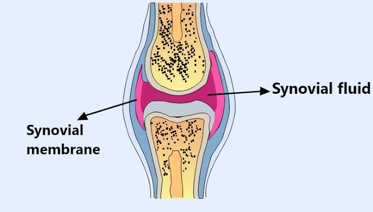
Synovial fluid (SF) is a critical component of joint health, serving multiple functions including lubrication, shock absorption, and nutrient distribution to the avascular cartilage. The biochemical composition of synovial fluid is complex and consists of several key components that work synergistically to maintain joint function and integrity.
1. Water Content
The primary constituent of synovial fluid is water, which makes up approximately 90-95% of its total volume. This high water content is essential for maintaining the fluid’s viscosity and enabling it to act as a lubricant within the joint.
2. Lubricants
Two major lubricating molecules found in synovial fluid are hyaluronan (HA) and lubricin (also known as proteoglycan 4).
- Hyaluronan (HA): This is a high-molecular-weight glycosaminoglycan that provides viscoelastic properties to the synovial fluid. HA contributes to the lubrication of articular cartilage surfaces, allowing for low-friction movement during joint motion.
- Lubricin: This glycoprotein plays a crucial role in reducing friction between cartilage surfaces. It also helps in protecting cartilage from wear and tear by forming a protective layer on the surface of the cartilage.
3. Proteins
Synovial fluid contains various proteins, including albumin and immunoglobulins, which are derived from blood plasma ultrafiltrate. These proteins serve several functions such as:
- Providing structural support
- Facilitating immune responses
- Contributing to the overall viscosity of the fluid
4. Electrolytes
Electrolytes such as sodium, potassium, calcium, and chloride are present in synovial fluid. These ions help maintain osmotic balance and contribute to the overall homeostasis within the joint environment.
5. Metabolic Byproducts
Synovial fluid also contains metabolic byproducts resulting from cellular metabolism within the joint space. These may include lactate and other organic acids that can provide insights into metabolic activity or pathological conditions affecting the joint.
6. Cytokines
Cytokines are signaling molecules that play an essential role in inflammation and immune responses within joints. Their presence in synovial fluid can indicate inflammatory processes associated with various arthritic conditions.
7. Crystals
In certain pathological conditions like gout or pseudogout, crystals may be present in synovial fluid due to abnormal metabolism of uric acid or calcium pyrophosphate dihydrate (CPPD). The identification of these crystals through microscopic analysis can aid in diagnosing specific types of arthritis.
Overall, changes in any component of synovial fluid can provide valuable diagnostic information regarding joint health or disease states such as infections, autoimmune disorders, or degenerative diseases.
Synthesis of Vitamin D-3
Introduction to Vitamin D-3 Synthesis
Vitamin D-3, also known as cholecalciferol, is synthesized in the body through a multi-step process that begins with the skin’s exposure to ultraviolet (UV) radiation. This synthesis is crucial for maintaining adequate levels of vitamin D, which plays an essential role in calcium metabolism and bone health.
Step 1: Formation of Previtamin D-3
The synthesis of vitamin D-3 starts when UVB rays from sunlight penetrate the skin and convert 7-dehydrocholesterol (a cholesterol derivative present in the skin) into previtamin D-3. This reaction occurs primarily in the epidermal layer of the skin. The chemical structure of 7-dehydrocholesterol allows it to absorb UV radiation effectively, leading to its transformation into previtamin D-3.
Step 2: Isomerization to Vitamin D-3
Once formed, previtamin D-3 undergoes a thermal isomerization process. This means that as it is exposed to heat (which can occur naturally from body temperature or environmental warmth), it rearranges its molecular structure to become vitamin D-3. This step is crucial because it converts the inactive form (previtamin D-3) into the active form (vitamin D-3).
Step 3: Conversion in the Liver
After its formation in the skin, vitamin D-3 enters the bloodstream and travels to the liver. In the liver, it undergoes hydroxylation—a chemical reaction where a hydroxyl group (-OH) is added—resulting in the formation of calcidiol (25-hydroxyvitamin D). Calcidiol is considered a storage form of vitamin D and serves as an important marker for assessing vitamin D status in individuals.
Step 4: Activation in the Kidneys
The final step occurs in the kidneys, where calcidiol is further hydroxylated to produce calcitriol (1,25-dihydroxyvitamin D), which is the biologically active form of vitamin D. Calcitriol plays a vital role in regulating calcium and phosphorus levels in the blood and promoting healthy bone formation.
Importance of Vitamin D-3 Synthesis
Vitamin D-3 synthesis is essential not only for bone health but also for overall immune function. Insufficient levels of vitamin D can lead to various health issues such as rickets in children and osteomalacia or osteoporosis in adults. Given that many people may not receive adequate sun exposure due to lifestyle or geographical factors, dietary sources and supplements are often recommended to maintain sufficient levels of this vital nutrient.
In summary, the synthesis of Vitamin D-3 involves several key steps: conversion of 7-dehydrocholesterol to previtamin D-3 via UV radiation, thermal isomerization to vitamin D-3, conversion to calcidiol in the liver, and finally activation to calcitriol in the kidneys.


