Regulation of Bone Modeling and Remodeling
Bone modeling and remodeling are crucial processes that maintain the integrity and functionality of the skeletal system. These processes are regulated by a complex interplay of mechanical, hormonal, and cellular factors.
Bone Modeling
Bone modeling refers to the process by which bones change shape or size in response to mechanical loads and physiological demands. This process is particularly important during growth periods, such as childhood and adolescence, when bones are still developing. The regulation of bone modeling involves:
- Mechanical Loading: The application of mechanical stress on bones stimulates osteocytes (mature bone cells) to communicate with osteoblasts (bone-forming cells) and osteoclasts (bone-resorbing cells). This communication leads to an increase in bone formation at sites experiencing higher loads while promoting resorption at less stressed areas.
- Cellular Communication: Osteocytes play a central role in sensing mechanical strain and orchestrating the activities of osteoblasts and osteoclasts through signaling pathways involving molecules like nitric oxide and prostaglandins.
- Hormonal Influence: Hormones such as parathyroid hormone (PTH), calcitonin, estrogen, and testosterone also influence bone modeling by modulating the activity of osteoblasts and osteoclasts.
Bone Remodeling
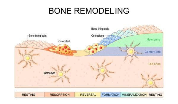
Bone remodeling is a continuous process that replaces old or damaged bone tissue with new bone without altering the overall shape of the skeleton. It consists of several phases:
- Quiescence: The resting phase where no significant cellular activity occurs.
- Activation: Signals from hormones or mechanical stress activate osteoclast precursors to differentiate into mature osteoclasts.
- Resorption: Osteoclasts resorb old bone tissue, creating micro-damage that signals for new bone formation.
- Reversal Phase: After resorption, there is a transition phase where the surface is prepared for new bone formation.
- Formation: Osteoblasts synthesize new bone matrix in response to signals from osteocytes and other factors.
- Mineralization: The newly formed matrix undergoes mineralization to become mature bone tissue.
- Termination: The remodeling cycle concludes when the activity ceases, returning to quiescence until the next cycle begins.
The balance between these phases is critical for maintaining healthy bone mass; disruptions can lead to conditions such as osteoporosis.
Systemic Regulation of Bone Modeling and Remodeling
The systemic regulation of bone modeling and remodeling involves various hormones, growth factors, and signaling pathways that coordinate these processes:
- Hormonal Regulators:
- Parathyroid Hormone (PTH): Secreted by the parathyroid glands, PTH increases blood calcium levels by stimulating osteoclastic activity while also promoting some aspects of osteoblastic function.
- Calcitonin (CT): Produced by the thyroid gland, calcitonin inhibits osteoclast activity, thereby reducing bone resorption.
- Vitamin D3 [1,25(OH)2 vitamin D3]: Enhances intestinal absorption of calcium and phosphate while also promoting both osteoclastic resorption and osteoblastic formation.
- Estrogen: Plays a protective role against excessive bone resorption; its deficiency can lead to increased risk for osteoporosis due to heightened osteoclastic activity.
- Growth Factors:
- Various growth factors such as Insulin-like Growth Factors (IGFs), Transforming Growth Factor-beta (TGF-β), Fibroblast Growth Factors (FGFs), Epidermal Growth Factor (EGF), WNT proteins, and Bone Morphogenetic Proteins (BMPs) significantly influence both modeling and remodeling processes by regulating cell proliferation, differentiation, survival, and matrix production.
- Cellular Interactions within Bone Multicellular Units (BMUs):
- BMUs consist of coordinated groups of cells including osteoblasts, osteoclasts, mesenchymal stem cells, and their precursors working together in a synchronized manner throughout the remodeling cycle.
- Communication among these cells through signaling molecules ensures that both formation and resorption occur effectively in response to physiological needs or external stimuli.
In summary, both bone modeling during growth phases and remodeling throughout life are tightly regulated processes influenced by mechanical forces as well as systemic hormonal signals that ensure optimal skeletal health.
Hormonal Control of Calcium
Calcium homeostasis is essential for various physiological functions, including bone health. The body regulates calcium levels through several hormones, primarily parathyroid hormone (PTH), calcitonin, and vitamin D. When serum calcium levels drop, PTH is released from the parathyroid glands, stimulating osteoclast activity to increase bone resorption and release calcium into the bloodstream. Conversely, when calcium levels are high, calcitonin is secreted from the thyroid gland to inhibit osteoclast activity and promote calcium deposition in bones.
Role of Vitamin D and Calcium Absorption
Vitamin D plays a crucial role in calcium absorption in the intestines. It enhances the intestinal absorption of dietary calcium by promoting the synthesis of calcium-binding proteins. The active form of vitamin D, calcitriol (1,25-dihydroxyvitamin D), increases the efficiency of calcium absorption significantly. Clinically, vitamin D deficiency can lead to conditions such as rickets in children or osteomalacia in adults due to impaired mineralization of bone.
The relationship between vitamin D and calcium absorption underscores its importance in maintaining adequate serum calcium levels for proper bone remodeling. Insufficient vitamin D can lead to secondary hyperparathyroidism as the body attempts to compensate for low serum calcium levels.
Role of Parathyroid Hormone
Parathyroid hormone (PTH) is a key regulator of bone remodeling. It is secreted by the parathyroid glands in response to low serum calcium levels. PTH acts on bones, kidneys, and intestines:
- Bones: PTH stimulates osteoclasts indirectly through osteoblasts, leading to increased bone resorption.
- Kidneys: PTH promotes renal tubular reabsorption of calcium while increasing phosphate excretion.
- Intestines: Although PTH does not act directly on the intestines, it enhances intestinal absorption of calcium indirectly via its effect on vitamin D metabolism.
Chronic elevation of PTH can lead to osteoporosis due to excessive bone resorption.
Synthesis and Regulation of Parathyroid Hormone
PTH is synthesized as a preprohormone in chief cells of the parathyroid glands. This preprohormone undergoes cleavage to form pro-PTH before being converted into active PTH (84 amino acids). The secretion of PTH is tightly regulated by serum calcium levels through a negative feedback mechanism: low serum calcium stimulates PTH release while high levels inhibit it.
Additionally, magnesium levels also influence PTH secretion; hypomagnesemia can stimulate PTH release while hypermagnesemia can suppress it.
Regulation of Calcitonin
Calcitonin is produced by parafollicular cells (C-cells) in the thyroid gland and serves as a counter-regulatory hormone to PTH. Its primary function is to lower blood calcium levels by inhibiting osteoclast activity and promoting renal excretion of calcium. Calcitonin secretion is stimulated by elevated serum calcium concentrations; however, its clinical significance appears less critical than that of PTH since many individuals can maintain normal physiology even with reduced calcitonin production.
Influence of Growth Hormone and Glucocorticoids
Growth hormone (GH) has anabolic effects on bone tissue; it stimulates chondrocyte proliferation at growth plates during childhood and adolescence leading to increased linear growth and skeletal mass. GH also promotes IGF-1 production from the liver which further stimulates osteoblast activity.
In contrast, glucocorticoids have catabolic effects on bone tissue; they inhibit osteoblast function while promoting apoptosis in these cells leading to decreased bone formation over time. Chronic exposure to glucocorticoids can result in significant bone loss known as glucocorticoid-induced osteoporosis.
In summary, systemic regulation of bone modeling and remodeling involves intricate hormonal control mechanisms primarily mediated by parathyroid hormone, calcitonin, vitamin D, growth hormone, and glucocorticoids that work together to maintain skeletal integrity throughout life.


