Anatomy of the Visual Field Pathway
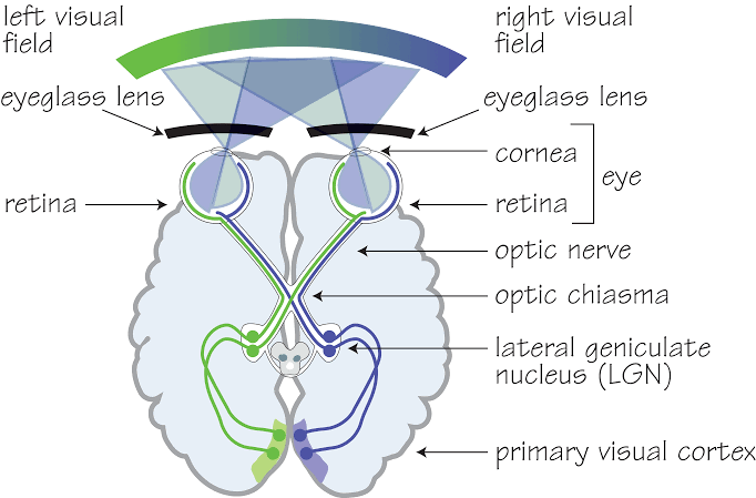
The visual field pathway is a complex system that transmits visual information from the retina to the brain, allowing for the perception of images. This pathway can be divided into several key anatomical structures, each playing a crucial role in processing visual stimuli.
1. Retina
The journey begins at the retina, which is the innermost layer of the eye. The retina contains photoreceptor cells known as rods and cones that detect light and convert it into electrical signals. Rods are more sensitive to light and are responsible for vision in low-light conditions, while cones are responsible for color vision and function best in bright light. The retina has ten layers, with bipolar cells acting as first-order neurons that receive signals from these photoreceptors.
2. Optic Nerve (CN II)
Once the photoreceptors have converted light into electrical impulses, these signals are transmitted to bipolar cells, which synapse with ganglion cells located in the inner layer of the retina. The axons of ganglion cells converge at the optic disc to form the optic nerve (cranial nerve II). This nerve carries both nasal and temporal fibers from one eye and is covered by meninges as it exits through the optic canal.
3. Optic Chiasm
As the optic nerves travel towards the brain, they meet at a midline structure called the optic chiasm. Here, nasal fibers from each eye decussate (cross over) while temporal fibers remain on their respective sides. This crossing allows visual information from both eyes to be processed together for depth perception and a unified field of view.
4. Optic Tract
After passing through the optic chiasm, visual information continues along the optic tract. Each optic tract contains fibers that transmit information about contralateral (opposite side) visual fields; thus, damage to one side of an optic tract can result in contralateral homonymous hemianopia (loss of half of the visual field on one side).
5. Lateral Geniculate Body (LGB)
The optic tract terminates at the lateral geniculate body (LGB), which is located in the posterior part of the thalamus. The LGB consists of six layers: layers 1, 4, and 6 receive input from nasal fibers while layers 2, 3, and 5 receive input from temporal fibers. The LGB acts as a relay station where further processing occurs before sending information to higher cortical areas.
6. Optic Radiation
From the LGB, visual signals travel via a white matter tract known as optic radiation or geniculocalcarine tract towards the primary visual cortex located in the occipital lobe. The optic radiation has two main pathways: some fibers pass directly through deep parts of the parietal lobe while others form Meyer’s loop around the inferior horn of lateral ventricle before reaching their destination.
7. Primary Visual Cortex
The primary visual cortex (Brodmann area 17) receives processed visual information via these pathways and is situated on both sides of the calcarine sulcus on the medial surface of each hemisphere’s occipital lobe. It registers information from contralateral visual fields—meaning that stimuli from one side are processed by the opposite hemisphere—and has distinct areas dedicated to central versus peripheral vision.
In summary, the anatomy of the visual field pathway involves multiple structures including:
- Retina
- Optic Nerve
- Optic Chiasm
- Optic Tract
- Lateral Geniculate Body
- Optic Radiation
- Primary Visual Cortex
These components work together seamlessly to ensure that visual stimuli are accurately perceived and interpreted by our brains.
Visual Field Defect in the Lesion of Optic Nerve
Lesions of the optic nerve can lead to significant visual field defects, primarily affecting the vision in one eye. The specific nature of these defects depends on the extent and location of the lesion within the optic nerve.
Complete Lesion of the Optic Nerve
When there is a complete lesion of the optic nerve, it results in total blindness in the affected eye. For instance, if there is damage to the right optic nerve, this will cause complete loss of vision in the right eye. This condition is known as unilateral blindness.
Partial Lesion of the Optic Nerve
In cases where only a portion of the optic nerve is affected, different types of visual field defects may occur:
- Central Scotoma: If the internal fibers of the optic nerve are involved, patients may experience a central scotoma. This defect manifests as a blind spot in their central vision while peripheral vision remains intact. Central scotomas can significantly impair tasks that require detailed vision, such as reading.
- Tunnel Vision: In cases where external fibers are affected (as seen in conditions like optic neuritis), patients may experience tunnel vision. This means that while central vision may be preserved to some degree, peripheral vision is severely restricted, leading to a “tunnel-like” view.
- Afferent Pupillary Defect: Patients with lesions in one optic nerve often exhibit an afferent pupillary defect (Marcus Gunn pupil). When light is shone into the affected eye, there is a relative decrease in constriction compared to when light is shone into the unaffected eye.
- Decreased Color Vision and Contrast Sensitivity: Lesions can also lead to decreased color perception and contrast sensitivity due to damage to specific pathways within the optic nerve.
Overall, lesions affecting different parts or fibers within the optic nerve can produce varying visual field defects ranging from complete blindness in one eye to more subtle changes like scotomas or tunnel vision.
The understanding of these visual field defects helps clinicians localize lesions based on physical examination and diagnostic testing.
Visual Field Defect in the Lesion of Optic Chiasma
The optic chiasm is a critical structure in the visual pathway where the optic nerves from both eyes partially cross. Lesions affecting the optic chiasm can lead to specific visual field defects, which are crucial for diagnosing and localizing the lesion. The most common visual field defect associated with optic chiasm lesions is bitemporal hemianopia. This defect results from the compression of the crossing fibers in the central part of the chiasm, leading to loss of the temporal visual fields in both eyes.
Types of Visual Field Defects
- Bitemporal Hemianopia: This is the classic visual field defect associated with optic chiasm lesions. It occurs when the central crossing fibers of the optic chiasm are compressed, leading to loss of the temporal visual fields in both eyes. This defect can be complete or partial, depending on the extent of the lesion.
- Junctional Scotoma: This defect occurs when the lesion is located at the junction of the optic nerve and the chiasm. It results in an ipsilateral central scotoma (loss of central vision in the eye on the same side as the lesion) and a contralateral superotemporal visual field defect. The junctional scotoma of Traquair is a variant where there is monocular hemianopic visual field loss.
- Chiasmal Bitemporal Hemianopsia Variants: Depending on the exact location and extent of the lesion, variations of bitemporal hemianopia can occur. For instance, a lesion affecting the posterior part of the chiasm might lead to a paracentral bitemporal hemianopsia due to the involvement of crossing macular fibers.
Diagnostic Criteria and Testing
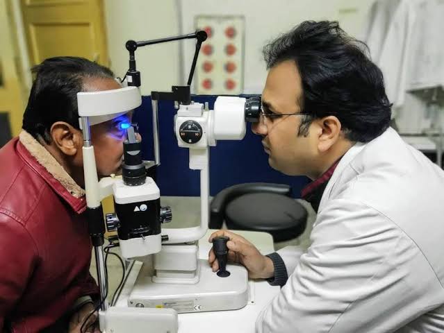
Visual field testing, such as the Humphrey visual field test, is essential for diagnosing optic chiasm lesions. According to what I know, a new criterion called the “simple temporal depression index” has been developed to assist in the diagnosis of compressive lesions of the optic chiasm. This index is calculated as the ratio of the sums of the thresholds for one line on the nasal side and temporal side of the vertical meridian. The sensitivity and specificity of this new criterion are reported to be 87% and 99%, respectively.
In addition, Fujimoto et al. proposed diagnostic criteria based on the Humphrey visual field test for pituitary adenomas, which are a common cause of optic chiasm compression. These criteria include:
- A vertical step, where at least four adjacent pairs of bilateral values on either side of the midline show diminished sensitivity of ≥2 dB on the temporal side, or at least three adjacent points with sensitivity diminished by ≥3 dB.
- A temporal depression index, where the difference between the nasal and temporal quadrant sums divided by the temporal quadrant sum exceeds specific thresholds (0.086 for the upper quadrant and 0.084 for the lower quadrant).
Etiology of Optic Chiasm Lesions
The most common cause of optic chiasm lesions is neoplastic in nature, with pituitary adenomas being the most frequent. Other neoplasms that can cause chiasmal compression include craniopharyngiomas, meningiomas (including optic nerve sheath meningiomas), and optic gliomas. Less common causes include chordomas, germinomas, endodermal sinus tumors, leukemias, lymphomas, nasopharyngeal carcinomas, and metastatic diseases. Non-neoplastic causes can also lead to chiasmal compression, such as sphenoid sinus mucoceles.
Conclusion
Lesions of the optic chiasm can lead to a variety of visual field defects, with bitemporal hemianopia being the most characteristic. Accurate diagnosis relies on detailed visual field testing and understanding the specific patterns of visual field loss associated with different types of lesions. The use of advanced diagnostic criteria and indices can enhance the accuracy of diagnosing and localizing optic chiasm lesions.
Diagnosis of Visual Field Defect in Post-Chiasmal Lesion
Understanding Post-Chiasmal Lesions
Post-chiasmal lesions refer to any pathological changes occurring after the optic chiasm along the visual pathway. These lesions can arise from various causes, including tumors, vascular issues, or traumatic injuries. The location of the lesion significantly influences the type of visual field defect observed.
Types of Visual Field Defects
- Homonymous Hemianopia: This is the most common visual field defect associated with post-chiasmal lesions. It occurs when there is damage to the optic tract, lateral geniculate nucleus (LGN), or optic radiations on one side of the brain. Patients typically experience loss of vision in either the right or left half of their visual field in both eyes.
- Quadrantanopia: In some cases, a post-chiasmal lesion may lead to quadrantanopia, which is a loss of vision in one quadrant (one-fourth) of the visual field. This can occur if there is damage localized to specific areas within the optic radiations.
- Scotomas: These are localized areas of vision loss that can occur due to lesions affecting specific parts of the visual pathway, such as those involving the occipital lobe where visual processing occurs.
Mechanism Behind Visual Field Defects
The mechanism behind these defects lies in how visual information is processed after it leaves the optic chiasm:
- Optic Tract Damage: If a lesion affects one optic tract, it results in homonymous hemianopia because all fibers carrying information from one half of each retina are disrupted.
- Optic Radiation Damage: Lesions affecting different segments of the optic radiations can lead to varying types of quadrantanopia depending on whether they affect superior or inferior fibers.
- Occipital Lobe Damage: Lesions in this area can cause more complex patterns such as scotomas or even complete loss depending on their extent and location.
Conclusion
In summary, post-chiasmal lesions typically result in homonymous hemianopia, but may also present as quadrantanopia or scotomas depending on their specific location and nature within the visual pathway.


