Overview of Iron Metabolism
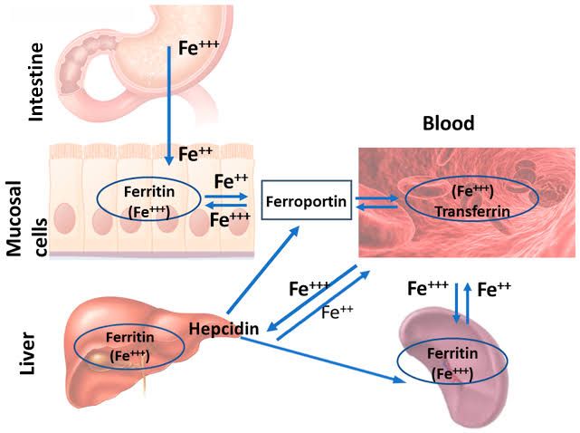
Iron metabolism is a complex process that involves the regulation of iron absorption, distribution, storage, and recycling within the body. The following steps outline the key components and mechanisms involved in iron metabolism:
1. Iron Absorption: Iron is primarily absorbed in the duodenum (the first part of the small intestine). There are two forms of dietary iron: heme iron (found in animal products) and non-heme iron (found in plant sources). Heme iron is more readily absorbed than non-heme iron. Non-heme iron must be reduced from its ferric (Fe^3+) to ferrous (Fe^2+) state for optimal absorption, a process facilitated by gastric acid and reducing agents like vitamin C.
Once reduced, ferrous iron is taken up by enterocytes (intestinal cells) through specific transporters such as divalent metal transporter 1 (DMT1). Inside enterocytes, iron can either be stored as ferritin or transported across the cell membrane into circulation.
2. Iron Export: The only known cellular exporter of iron is ferroportin (Fpn), which transports iron from enterocytes into the bloodstream. The release of iron into circulation is tightly regulated by hepcidin, a peptide hormone produced by the liver. Hepcidin binds to ferroportin, leading to its internalization and degradation, thereby decreasing serum iron levels.
3. Transport in Blood: Once released into circulation, ferrous iron is oxidized back to ferric form and binds to transferrin, a plasma protein that transports iron throughout the body. Transferrin-bound iron is then delivered to various tissues where it is needed for processes such as hemoglobin synthesis in erythrocytes (red blood cells).
4. Iron Utilization: Cells take up transferrin-bound iron via transferrin receptors (TfR), which are particularly abundant on cells with high demand for iron, such as erythroid progenitor cells in the bone marrow. Once inside the cell, transferrin-iron complexes are internalized through endocytosis and released from endosomes for use in various cellular functions.
5. Iron Storage: Excess iron that is not immediately needed for biological functions is stored primarily in hepatocytes (liver cells) as ferritin or hemosiderin. Ferritin serves as a readily accessible source of stored iron that can be mobilized when needed.
6. Iron Recycling: Iron recycling occurs mainly through macrophages that phagocytose senescent red blood cells. These macrophages break down hemoglobin to release heme, which is further degraded to release free iron. This recycled iron can then be reused for new erythrocyte production or stored again.
7. Regulation of Iron Homeostasis: The regulation of systemic iron homeostasis involves several key players:
- Hepcidin: The master regulator that controls serum iron levels by modulating ferroportin activity.
- Ferroportin: The primary exporter responsible for releasing intracellularly stored or absorbed iron into circulation.
- Transferrin: The main transport protein that carries ferric ions throughout the bloodstream.
- Transferrin Receptors: Facilitate cellular uptake of transferrin-bound iron.
Dysregulation of these components can lead to disorders such as hereditary hemochromatosis (iron overload) or anemia due to insufficient available iron.
Iron Metabolism: Mechanism of Absorption and Factors Affecting It
Mechanism of Iron Absorption
Iron absorption primarily occurs in the duodenum and upper jejunum of the small intestine. There are two forms of dietary iron: heme iron (found in animal products) and non-heme iron (found in plant-based foods).
- Heme Iron Absorption: Heme iron is absorbed more efficiently than non-heme iron. It enters enterocytes (intestinal cells) via specific transporters, although the exact mechanism remains less understood.
- Non-Heme Iron Absorption: Non-heme iron must be reduced from its ferric form (Fe3+) to ferrous form (Fe2+) before absorption. This reduction is facilitated by gastric acid and enzymes like duodenal cytochrome B reductase (DCYTB). Once reduced to Fe2+, it is taken up by enterocytes through the apical divalent metal transporter 1 (DMT1).
- Intracellular Handling: Inside enterocytes, non-utilized iron can be stored as ferritin or exported into circulation via ferroportin, a basolateral membrane protein. The export process is regulated by hepcidin, a peptide hormone produced by the liver that controls systemic iron levels.
- Transport in Blood: Once released into circulation, ferrous iron is oxidized back to ferric form and binds to transferrin, a plasma protein that transports iron to various tissues.
Factors Affecting Iron Absorption
Several factors influence the efficiency of iron absorption:
- Dietary Composition:
- Enhancers: Vitamin C (ascorbic acid) can enhance non-heme iron absorption by reducing ferric to ferrous form and forming soluble complexes.
- Inhibitors: Compounds such as phytates (found in grains), polyphenols (in tea and coffee), calcium, and certain proteins can inhibit non-heme iron absorption by binding to it or forming insoluble complexes.
- Iron Status:
- Individuals with low body iron stores exhibit increased intestinal absorption due to enhanced expression of DMT1 and reduced hepcidin levels.
- Conversely, those with high body iron stores experience decreased absorption due to elevated hepcidin levels which inhibit ferroportin activity.
- Physiological Conditions:
- Conditions such as pregnancy or growth spurts increase the body’s demand for iron, leading to enhanced absorption.
- Inflammatory states can elevate hepcidin levels due to cytokines like interleukin-6, resulting in reduced intestinal absorption despite adequate dietary intake.
- Age and Gender:
- Pre-menopausal women typically have higher daily losses due to menstruation which increases their dietary requirements for iron.
- Infants and children have higher needs during growth phases which may necessitate increased dietary intake.
- Gastrointestinal Health:
- Disorders affecting gut health such as celiac disease or inflammatory bowel disease can impair nutrient absorption including that of iron.
In summary, effective regulation of dietary intake alongside understanding individual physiological conditions plays a crucial role in maintaining optimal iron metabolism within the body.
Interpretation of Iron Deficiency Anemia Based on Given Data and Microscopic Findings
Definition and Diagnosis
Iron deficiency anemia (IDA) is characterized by a decrease in hemoglobin levels below the normal range, alongside laboratory findings indicative of iron deficiency. The diagnosis is confirmed through specific tests that include serum ferritin, serum iron, total iron-binding capacity (TIBC), and transferrin saturation. A low ferritin level is diagnostic for IDA, while in cases of coexisting inflammation or malignancy, ferritin levels may be misleadingly elevated.
Laboratory Findings
- Ferritin Levels: Ferritin serves as the primary indicator of iron stores in the body. In IDA, ferritin levels are typically low (<20 μg/L). If ferritin is between 20-100 μg/L, further investigation is warranted to rule out other conditions such as anemia of chronic disease (ACD).
- Serum Iron and TIBC: In IDA, serum iron levels are low, while TIBC is elevated. This reflects the body’s compensatory mechanism to increase iron transport due to low availability.
- Transferrin Saturation: The transferrin saturation percentage will also be low in IDA due to reduced serum iron levels.
- Mean Cell Volume (MCV) and Mean Cell Hemoglobin (MCH): Both MCV and MCH are typically decreased in IDA, indicating microcytic and hypochromic red blood cells.
- Peripheral Blood Smear: Microscopic examination reveals microcytic (smaller than normal) and hypochromic (less color than normal) red blood cells. Anisocytosis (variation in RBC size) may also be present.
Clinical Symptoms
Patients with IDA often report symptoms such as fatigue, pallor, cold intolerance, leg cramps during exertion, cravings for non-nutritive substances like ice or dirt (pica), and impaired cognitive function in children. Physical examination may reveal signs like pallor of mucous membranes, spoon-shaped nails (koilonychia), glossitis (glossy tongue), angular stomatitis (fissures at the corners of the mouth), and splenomegaly in severe cases.
Management
The management of IDA involves identifying the underlying cause—such as dietary deficiencies, malabsorption syndromes like celiac disease, or gastrointestinal bleeding—and addressing it accordingly. Iron supplementation through oral or intravenous routes is commonly employed to replenish iron stores. Regular monitoring of hemoglobin and ferritin levels post-treatment is essential to ensure effective recovery.
In summary, interpreting iron deficiency anemia requires a comprehensive approach involving clinical evaluation supported by laboratory findings that confirm low iron stores and altered red blood cell morphology consistent with microcytic anemia.
Interpretation of Folic Acid and Cobalamin in Relation to Anemias
Overview of Megaloblastic Anemia (MBA)
Megaloblastic anemia is characterized by the presence of large, abnormal red blood cells (RBCs) known as megaloblasts, which arise due to impaired DNA synthesis. This condition is primarily caused by deficiencies in vitamin B12 (cobalamin) and vitamin B9 (folate), leading to ineffective erythropoiesis and increased apoptosis in developing erythrocytes.
Role of Folic Acid (Vitamin B9)
Folic acid is crucial for DNA synthesis, repair, and methylation processes. In the context of megaloblastic anemia, a deficiency in folate results in the inhibition of thymidylate synthesis, which is essential for DNA replication. This inhibition leads to the accumulation of deoxyuridine monophosphate (dUMP), causing DNA damage and ultimately resulting in cell death or apoptosis. The information provided indicates that 88% of patients with MBA had low levels of folate, correlating with elevated homocysteine levels. High homocysteine can further contribute to neurological issues and cardiovascular diseases.
Role of Cobalamin (Vitamin B12)
Cobalamin plays a vital role in the metabolism of fatty acids and amino acids, as well as in the production of myelin sheaths around nerves. A deficiency in vitamin B12 can lead to similar disruptions in DNA synthesis as seen with folate deficiency. Specifically, it affects the conversion of methylmalonyl-CoA to succinyl-CoA and impacts the formation of tetrahydrofolate (THF), which is necessary for purine synthesis. In this study, 80% of MBA patients exhibited low cobalamin levels, indicating a significant correlation between vitamin B12 deficiency and megaloblastic anemia.
Microscopic Findings
Microscopically, bone marrow biopsies from patients with megaloblastic anemia reveal hypercellularity with an abundance of megaloblasts—large nucleated cells that are indicative of impaired maturation due to vitamin deficiencies. The immunohistochemical analysis showed elevated p53 expression in these samples compared to control subjects without MBA. The high levels of p53 suggest that apoptosis is being induced due to the inability to repair DNA damage effectively caused by low folate and cobalamin levels.
Conclusion
In summary, both folic acid and cobalamin deficiencies play critical roles in the pathogenesis of megaloblastic anemia through their involvement in DNA synthesis and cellular maturation processes. Their deficiencies lead to ineffective erythropoiesis characterized by large immature RBCs that undergo apoptosis rather than maturing into functional erythrocytes.
Biochemical Role of Pyridoxine and Vitamin C in Microcytic Anemia
Introduction to Microcytic Anemia
Microcytic anemia is characterized by the presence of smaller-than-normal red blood cells (RBCs) and is often associated with iron deficiency, thalassemia, or chronic disease. The biochemical roles of vitamins such as pyridoxine (vitamin B6) and vitamin C are crucial in the context of this condition.
Role of Pyridoxine (Vitamin B6)
Pyridoxine plays a significant role in hemoglobin synthesis and the overall metabolism of amino acids. It is a coenzyme for various enzymatic reactions involved in the production of neurotransmitters and the synthesis of heme, which is an essential component of hemoglobin.
- Heme Synthesis: Pyridoxine is vital for the synthesis of 5-aminolevulinic acid (ALA), a precursor to porphyrin, which eventually leads to heme formation. A deficiency in pyridoxine can impair heme production, leading to reduced hemoglobin levels and contributing to microcytic anemia.
- Amino Acid Metabolism: Pyridoxine is involved in transamination reactions that convert amino acids into different forms necessary for protein synthesis and energy metabolism. This function supports erythropoiesis (the production of red blood cells) by ensuring that adequate building blocks are available for hemoglobin synthesis.
- Immune Function: Adequate levels of pyridoxine are also important for maintaining immune function, which can be compromised in individuals with anemia, further complicating their health status.
Role of Vitamin C
Vitamin C (ascorbic acid) has several critical functions that impact microcytic anemia:
- Iron Absorption: One of the most notable roles of vitamin C is its ability to enhance non-heme iron absorption from plant-based sources. It reduces ferric iron (Fe^3+) to ferrous iron (Fe^2+), which is more readily absorbed in the intestines. This increased bioavailability can help address iron deficiency, a common cause of microcytic anemia.
- Antioxidant Properties: Vitamin C acts as an antioxidant, protecting cells from oxidative stress that can damage red blood cells and other components involved in hematopoiesis (the formation of blood cellular components). By reducing oxidative stress, vitamin C helps maintain healthy RBCs.
- Collagen Synthesis: Vitamin C is essential for collagen synthesis, which plays a role in maintaining the integrity of blood vessels and supporting overall circulatory health. Healthy vasculature ensures efficient delivery of nutrients and oxygen to tissues, including those involved in erythropoiesis.
- Regeneration of Other Antioxidants: Vitamin C helps regenerate other antioxidants like vitamin E, further enhancing its protective role against oxidative damage within the body.
Conclusion
In summary, both pyridoxine and vitamin C play integral roles in addressing microcytic anemia through their involvement in heme synthesis, amino acid metabolism, iron absorption enhancement, antioxidant protection, and overall support for erythropoiesis. Ensuring adequate intake of these vitamins may be beneficial for individuals suffering from this type of anemia.


