Derivatives of Fetal Vessels and Structures
1. Umbilical Vein
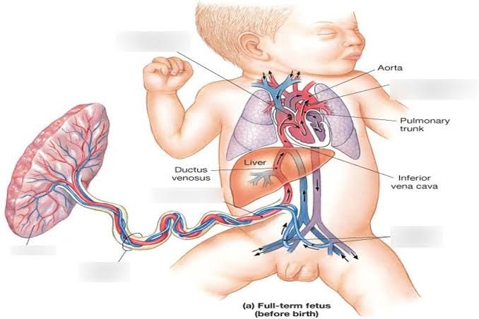
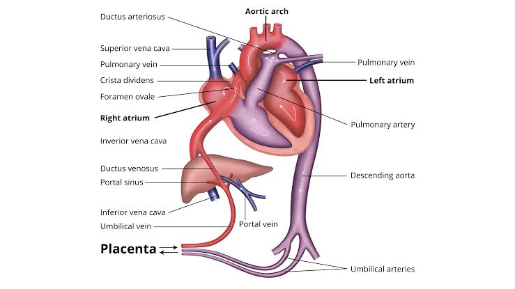
The umbilical vein is responsible for carrying oxygenated blood from the placenta to the fetus. After birth, it undergoes a transformation:
- Derivative: Ligamentum teres (round ligament of the liver). The umbilical vein obliterates and becomes a fibrous cord that runs along the free edge of the falciform ligament.
2. Ductus Venosus
The ductus venosus is a shunt that allows blood to bypass the liver, directing it into the inferior vena cava. After birth, this structure also changes:
- Derivative: Ligamentum venosum. The ductus venosus closes and becomes a fibrous band that connects the left branch of the portal vein to the inferior vena cava.
3. Umbilical Artery
The umbilical arteries carry deoxygenated blood from the fetus back to the placenta. Following delivery, these vessels are transformed:
- Derivative: Medial umbilical ligaments. The umbilical arteries constrict and eventually become fibrous cords that run along the lateral aspects of the bladder.
4. Foramen Ovale
The foramen ovale is an opening between the right and left atria of the fetal heart, allowing blood to bypass pulmonary circulation. After birth, it typically closes:
- Derivative: Fossa ovalis. The foramen ovale becomes a depression in the interatrial septum as it closes.
5. Ductus Arteriosus
The ductus arteriosus connects the pulmonary artery to the descending aorta, allowing blood to bypass non-functioning fetal lungs. After birth, this vessel also undergoes change:
- Derivative: Ligamentum arteriosum. The ductus arteriosus constricts and eventually forms a fibrous remnant connecting the pulmonary artery to the aorta.
Fetal and Neonatal Circulation
Fetal Circulation
In the fetus, the primary organ for gas exchange is the placenta, not the lungs, which are filled with liquid and do not function in gas exchange. The fetal circulatory system is designed to efficiently deliver oxygenated blood from the placenta to the systemic organs and return deoxygenated blood to the placenta for reoxygenation. This is achieved through a series of shunts and specific blood flow patterns.
- Oxygenated Blood Pathway: Oxygenated blood from the placenta travels through the umbilical vein to the ductus venosus, which directs this blood into the inferior vena cava (IVC). The ductus venosus and the Eustachian valve within the right atrium help to direct most of this oxygenated blood across the foramen ovale into the left atrium. From the left atrium, it enters the left ventricle and is pumped into the ascending aorta, supplying the upper body and brain with oxygen-rich blood.
- Deoxygenated Blood Pathway: Deoxygenated blood from the lower body returns to the right atrium via the IVC. This blood mixes minimally with the oxygenated blood due to the anatomical arrangement and flows into the right ventricle. The right ventricle then pumps this blood into the pulmonary artery. However, due to high pulmonary vascular resistance (PVR) in the fetus, most of this blood bypasses the lungs and flows through the ductus arteriosus (DA) into the descending aorta, where it mixes with some oxygenated blood from the ascending aorta and is directed to the lower body and back to the placenta for reoxygenation.
- Shunts and Flows: The key shunts in fetal circulation are the ductus venosus, the foramen ovale, and the ductus arteriosus. These shunts allow for the efficient distribution of blood to the systemic organs while bypassing the non-functional fetal lungs.
Neonatal Circulation
At birth, the transition from fetal to neonatal circulation involves significant physiological changes to adapt to the new environment where the lungs become the primary organ for gas exchange.
- Closure of Shunts: The first breath and subsequent lung expansion lead to a decrease in PVR, allowing more blood to flow into the lungs. This change in PVR, along with the clamping of the umbilical cord, increases systemic vascular resistance (SVR). These changes promote the closure of the foramen ovale due to increased left atrial pressure, and the ductus arteriosus begins to constrict due to increased oxygen levels and decreased prostaglandin levels.
- Establishment of Neonatal Circulation: With the closure of the fetal shunts, the neonatal circulation is established. Oxygenated blood from the lungs enters the left atrium, passes into the left ventricle, and is pumped into the systemic circulation via the aorta. Deoxygenated blood from the systemic circulation returns to the right atrium, enters the right ventricle, and is pumped into the pulmonary circulation via the pulmonary artery.
Transitional Neonatal Circulation
The transition from fetal to neonatal circulation is a critical period that can vary significantly between infants, particularly between term and preterm infants. This transitional phase involves the gradual closure of fetal shunts and the adaptation of the cardiovascular system to the postnatal environment.
- Physiological Changes: During this transition, there are alterations in preload, contractility, and afterload. The infant’s cardiovascular system must adapt to the increased oxygen levels, the shift from placental to pulmonary gas exchange, and the closure of the ductus arteriosus and foramen ovale.
- Clinical Implications: Understanding the physiology of transitional circulation is crucial for managing infants with hemodynamic compromise. Common issues during this period include patent ductus arteriosus (PDA), hypotension, intraventricular hemorrhage, birth asphyxia, severe growth restriction, and pulmonary hypertension. These conditions can lead to circulatory failure if not managed appropriately.
- Monitoring and Management: Advanced hemodynamic monitoring tools, such as echocardiography, near-infrared spectroscopy (NIRS), and electrical velocimetry, are increasingly used at the bedside to assess the cardiovascular status of neonates during this transition. Intact umbilical cord resuscitation is also a practice that can aid in the smooth transition of circulation.
- Impact on Health Outcomes: The objective assessment of cardio-respiratory transition and understanding of physiology in both normal and disease states can improve short- and long-term health outcomes for neonates. Proper management during this critical period can prevent complications and ensure a successful transition to neonatal circulation.
Clinical Implications of Transitional Neonatal Circulation
The clinical implications of transitional neonatal circulation are significant, as they directly impact the management and outcomes of newborns, especially those with congenital heart disease or preterm infants.
- Congenital Heart Disease: For infants with congenital heart defects, the fetal shunt pathways can redistribute ventricular blood flows to maintain adequate systemic blood flow during fetal life. However, after birth, the closure of these shunts can lead to circulatory compromise. For example, infants with severe left heart obstruction rely on the ductus arteriosus for systemic blood flow, and its closure can lead to circulatory failure. Similarly, infants with severe right heart obstruction depend on the ductus arteriosus for pulmonary blood flow, and its closure can result in critically low pulmonary blood flow.
- Preterm Infants: Preterm infants have an immature circulation, which can complicate the transition. Conditions such as PDA are more common in preterm infants and can lead to significant hemodynamic instability. The management of these infants requires a thorough understanding of the transitional circulation to prevent complications such as intraventricular hemorrhage and chronic lung disease.
- Management Strategies: The use of prostaglandin E1 (PGE1) to maintain the patency of the ductus arteriosus in infants with ductal-dependent lesions is a critical intervention during the transitional period. Additionally, the use of advanced monitoring techniques and understanding the physiological changes during transition can guide clinicians in making informed decisions to manage hemodynamic compromise effectively.
In conclusion, the transition from fetal to neonatal circulation is a complex process that requires careful monitoring and management to ensure a successful adaptation to postnatal life. Understanding the physiological changes and clinical implications of this transition is essential for improving the outcomes of newborns, particularly those with congenital heart disease or preterm infants.


