Types of Nerve Injury
There are three main types of nerve injury classified based on the severity and nature of the damage:
- Neuropraxia: This type involves a physiological block of nerve conduction within an axon without any anatomical interruption. Neuropraxia is often temporary, with recovery typically occurring within 4 to 6 weeks.
- Axonotmesis: This injury is characterized by an anatomical interruption of the axon while the connective tissue framework remains intact or only partially disrupted. Recovery requires regrowth of the axon to its target muscle, which can take a considerable amount of time and may be affected by scar formation.
- Neurotmesis: This is the most severe type of nerve injury, involving complete anatomical disruption of both the axon and surrounding connective tissue. Neurotmesis has no chance for spontaneous recovery, and early surgical intervention is necessary for potential recovery.
Wallerian Degeneration
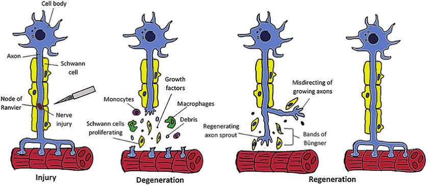
Wallerian degeneration is a biological process that occurs following a nerve injury, specifically when an axon is severed or damaged. This process involves the degeneration of the distal segment of the axon, which occurs in an anterograde manner, meaning it progresses away from the cell body towards the nerve terminal. The degeneration typically begins within 30 minutes to several days after the injury and can last for weeks.
Stages of Wallerian Degeneration
The process of Wallerian degeneration can be divided into three main stages:
- Axon Degeneration:
Shortly after injury, there is a separation between the proximal (nearer to the cell body) and distal (farther from the cell body) ends of the axon. The membranes at these ends seal off initially, but soon thereafter, degeneration begins. This stage is characterized by the formation of axonal sprouts that allow for potential nerve regeneration. - Myelin Clearance:
By around day seven post-injury, macrophages are recruited to clear away myelin debris and other cellular remnants. This cleanup is facilitated by Schwann cells, which play a crucial role in signaling macrophages to perform phagocytosis on degenerated myelin and axonal components. The rate of clearance differs significantly between the Peripheral Nervous System (PNS) and Central Nervous System (CNS), with PNS generally exhibiting faster clearance due to differences in blood-tissue barrier permeability and cellular responses. - Regeneration:
If the neuron’s cell body remains intact, regeneration can occur at a rate of approximately 1 mm per day. During this phase, cytoplasmic elements accumulate at the site of injury as granular disintegration takes place over several days to weeks.
Clinical Presentation
Patients experiencing Wallerian degeneration may present with various symptoms depending on which nerves are affected. Common clinical manifestations include:
- Reduced or loss of function in structures associated with damaged nerves.
- Numbness or tingling sensations that may spread from extremities upward.
- Sharp or burning pain.
- Increased sensitivity to touch.
- Muscle weakness or paralysis if motor nerves are involved.
- Coordination issues leading to falls.
Diagnostic Procedures
Diagnosis often involves several tests including:
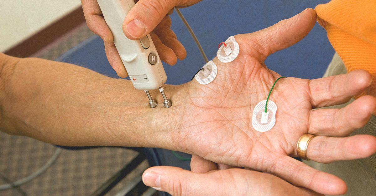
- Electromyography (EMG)
- Nerve conduction studies
- Assessments for sensory deficits
- Evaluations of muscle strength
These diagnostic tools help determine the extent of nerve damage and guide treatment options.
Management and Interventions
Management strategies for Wallerian degeneration focus on promoting nerve repair and recovery. These may include:
- Cryotherapy
- Exercise regimens
- Neurorehabilitation techniques
- Surgical interventions if necessary
The effectiveness of recovery largely depends on how well these interventions align with the innate immune response triggered by Wallerian degeneration.
In summary, Wallerian degeneration is a critical physiological response following peripheral nerve injury that facilitates both clearance of debris and potential regeneration of damaged nerves.
Regeneration of Nerve Fibers
The process of nerve fiber regeneration primarily occurs in the peripheral nervous system (PNS) and involves several key steps that facilitate recovery after injury. Here’s a detailed breakdown of this complex process:
1. Initial Response to Injury
When a peripheral nerve is injured, the immediate response involves the degeneration of the distal segment of the axon, known as Wallerian degeneration. This process begins when the axon is severed, leading to the breakdown of myelin sheaths surrounding the distal segment. The proximal segment may either undergo apoptosis (programmed cell death) or initiate a chromatolytic reaction, which is an attempt at repair.
2. Clearance of Debris
Following injury, there is a rapid migration of phagocytes, Schwann cells, and macrophages to the site of damage. These cells play a crucial role in clearing away cellular debris and damaged tissue resulting from Wallerian degeneration. This cleanup process is essential for creating an environment conducive to regeneration.
3. Schwann Cell Activation and Axonal Sprouting
Once debris is cleared, Schwann cells become activated and proliferate at the injury site. They form a supportive environment for regenerating axons by secreting neurotrophic factors that promote growth and survival. The proximal end of the severed axon begins to sprout new axonal processes in an effort to reconnect with target tissues.
4. Formation of Regeneration Tubes
Schwann cells align themselves along the path where the original nerve fibers were located, forming structures known as regeneration tubes or bands of Büngner. These tubes guide the growing axons toward their target tissues and provide structural support during regeneration.
5. Remyelination
As regenerating axons grow through these tubes, they begin to remyelinate with Schwann cell support. This remyelination is critical for restoring proper conduction velocities along the regenerated fibers, allowing for effective signal transmission.
6. Functional Recovery
The success of nerve fiber regeneration can lead to varying degrees of functional recovery depending on factors such as the type and extent of injury, age, health status of the individual, and timely intervention following injury. In optimal conditions, some functional recovery can be achieved; however, clinical outcomes can often be disappointing due to complications such as improper alignment or misdirection during regrowth.
In summary, the regeneration of nerve fibers in the PNS involves initial degeneration followed by clearance of debris, activation and proliferation of Schwann cells, axonal sprouting guided by regeneration tubes formed by Schwann cells, remyelination of newly formed axons, and ultimately functional recovery, which may vary based on several influencing factors.
Causes, Features, and Pathophysiology of Multiple Sclerosis and Guillain-Barré Syndrome
Causes
Multiple Sclerosis (MS) and Guillain-Barré Syndrome (GBS) are both autoimmune diseases that affect the nervous system, but they have different triggers and underlying causes.
- Multiple Sclerosis (MS):
- The exact cause of MS is not fully understood, but several factors may contribute to its development:
- Genetic Factors: There is a genetic predisposition to MS, as it tends to run in families.
- Environmental Factors: Certain environmental factors such as geographic location, exposure to sunlight (which affects vitamin D levels), and viral infections (notably the Epstein-Barr virus) are thought to play a role.
- Lifestyle Factors: Smoking has been identified as a risk factor for developing MS.
- The exact cause of MS is not fully understood, but several factors may contribute to its development:
- Guillain-Barré Syndrome (GBS):
- GBS often occurs following an infection. Common triggers include:
- Infections: Viral or bacterial infections such as those caused by Campylobacter jejuni, cytomegalovirus, Epstein-Barr virus, Zika virus, or influenza can precede GBS.
- Vaccinations: In rare cases, GBS has been reported after vaccinations.
- The immune response triggered by these infections may mistakenly attack the peripheral nerves.
- GBS often occurs following an infection. Common triggers include:
Features
- Multiple Sclerosis (MS):
- Symptoms of MS can vary widely but commonly include:
- Weakness
- Numbness or tingling in limbs
- Bladder dysfunction
- Fatigue
- Dizziness
- Muscle stiffness or spasms
- Vision problems (such as blurred vision)
- MS symptoms can be episodic with periods of exacerbation followed by remission.
- Symptoms of MS can vary widely but commonly include:
- Guillain-Barré Syndrome (GBS):
- GBS typically presents with rapid onset symptoms that may include:
- Weakness starting in the legs and progressing upwards
- Numbness or tingling sensations
- Difficulty walking
- Severe muscle weakness that can lead to paralysis
- Unlike MS, GBS symptoms usually peak within weeks and then gradually improve over time.
- GBS typically presents with rapid onset symptoms that may include:
Pathophysiology
- Multiple Sclerosis (MS):
- In MS, the immune system attacks myelin—the protective sheath surrounding nerve fibers in the central nervous system (CNS). This demyelination disrupts communication between the brain and other parts of the body.
- The process involves T cells crossing the blood-brain barrier and attacking oligodendrocytes (the cells that produce myelin), leading to inflammation and lesions in the CNS.
- Over time, this damage can result in scar tissue formation (sclerosis), which contributes to neurological deficits.
- Guillain-Barré Syndrome (GBS):
- In GBS, the immune system mistakenly targets peripheral nerves after an infection. This leads to demyelination of peripheral nerves.
- The pathophysiological mechanism often involves molecular mimicry where antibodies generated against infectious agents cross-react with nerve components.
- This results in inflammation and damage to myelin sheaths in peripheral nerves, leading to impaired nerve conduction and muscle weakness.
In summary, while both MS and GBS are autoimmune disorders characterized by demyelination of nerve tissues, they differ significantly in their causes, clinical features, affected nervous systems (central vs. peripheral), and pathophysiological mechanisms.


